Інформація призначена тільки для фахівців сфери охорони здоров'я, осіб,
які мають вищу або середню спеціальну медичну освіту.

Підтвердіть, що Ви є фахівцем у сфері охорони здоров'я.

Журнал «Актуальная инфектология» Том 8, №5, 2020

Вернуться к номеру

Клінічний випадок і літературний огляд патогенетичних аспектів комбінованої інфекції вірусу Епштейна — Барр та кандидозу порожнини рота

Авторы: S. Nykytyuk, S. Levenets, T. Kosovska, T. Nedoshytko
I. Horbachevsky Ternopil National Medical University, Ternopil, Ukraine

Рубрики: Инфекционные заболевания

Разделы: Справочник специалиста

Версия для печати


Резюме

Актуальність. Лікування пацієнтів iз рецидивуючою інфекцією, стійкою до традиційної терапії, є складним клінічним завданням у педіатрії. Мета роботи: проаналізувати існуючу літературу щодо клінічних особливостей кандидозу порожнини рота і інфекційного мононуклеозу. Матеріали та методи. Описаний випадок комбінованої вірусної інфекції Епштейна — Барр у формі інфекційного мононуклеозу і кандидозу ротової порожнини рота в 4-річного хлопчика. Результати. Досліджено бактеріальну флору горла і рота, в культурах iз горла були ізольовані Str.viridans 103 КУО/мл, S.aureus 105 КУО/мл, Candida albicans 106 КУО/мл. Імуноферментний аналіз показав, що індекс антитіл EBV VCA IgM становив 2,63 (більше 0,8 — позитивний результат). Висновки. При інфекційному мононуклеозі в поєднанні з кандидозом порожнини рота спостерігаються такі загальні симптоми: тривала лихоманка, лімфаденопатія і синдром екзантеми. Зазвичай вони виникають після прийому антибактеріальних засобів широкого спектра дії. У разі захворювання з такими симптомами, як тривала лихоманка, лімфаденопатія, тонзиліт і синдром екзантеми, що є спільними для кандидозу порожнини рота та інфекційного мононуклеозу, потрібна диференційна діагностика. Необхідно вивчити мікробіоту ротоглотки в пацієнтів з інфекційним мононуклеозом для виявлення флори, що може обумовлювати ускладнений перебіг захворювання і труднощі діагностики. У такому випадку цей вид дослідження є важливим діагностичним методом виявлення кандидозу.

Актуальность. Лечение пациентов с рецидивирующей инфекцией, которая устойчива к традиционной терапии, является сложной клинической задачей в педиатрии. Цель работы: проанализировать существующую литературу о клинических особенностях кандидоза полости рта и инфекционного мононуклеоза. Материалы и методы. Представлен случай комбинированной вирусной инфекции Эпштейна — Барр в форме инфекционного мононуклеоза и кандидоза ротовой полости рта у 4-летнего мальчика. Результаты. Исследована бактериальная флора горла и рта, в культурах из горла были изолированы Str.viridans 103 КОЕ/мл, S.aureus 105 КОЕ/мл, Candida albicans 106 КОЕ/мл. Иммуноферментный анализ показал, что индекс антител EBV VCA IgM составлял 2,63 (более 0,8 — положительный результат). Выводы. При инфекционном мононуклеозе в сочетании с кандидозом полости рта наблюдаются следующие общие симптомы: длительная лихорадка, лимфаденопатия и синдром экзантемы. Обычно они возникают после приема антибактериальных средств широкого спектра действия. В случае заболевания с такими симптомами, как длительная лихорадка, лимфаденопатия, тонзиллит и синдром экзантемы, которые являются общими для кандидоза полости рта и инфекционного мононуклеоза, требуется дифференциальная диагностика. Необходимо изучить микробиоту ротоглотки у пациентов с инфекционным мононуклеозом для выявления флоры, которая может обусловливать осложненное течение заболевания и трудности диагностики. В таком случае данный вид исследования является важным диагностическим методом обнаружения кандидоза.

Background. Patients with a recurrent infection that is resistant to traditional therapy are a clinical challenge in pediatrics. This recurrent pathology is often hiding another disease. In addition, timely diagnosis and suggestion of etiotropic therapy are often delayed when medical ethics have been deviated, namely, when medical history collection was dubious. The objective of this article was to present the clinical peculiarities of the Epstein-Barr virus (EBV) association with oral candidiasis. Materials and methods. The authors describe the clinical case of Epstein-Barr viral infection in a 4-year-old child. The case was detected in Ternopil region. Results. The bacterial flora of the throat and mouth was inoculated, in the throat culture, Str.viridans 103 CFU/ml, S.aureus 105 CFU/ml, Candida albicans 106 CFU/ml were isolated. Enzyme-linked immunoassay showed that EBV VCA IgM antibody index was at the level of 2.63 (more than 0.8 — a positive result). Conclusions. When infectious mononucleosis is combined with oral candidiasis, the following common symptoms are observed: prolonged fever, lymphadenopathy, and exanthema syndrome. They usually occur after administration of broad-spectrum antibacterial agents. In case of disease with symptoms such as prolonged fever, lymphadenopathy, tonsillitis, and exanthema syndrome, which are common for both oral candidiasis and infectious mononucleosis, differential diagnosis is required. There is a need to examine the microbiota of the oropharynx in patients with infectious mononucleosis to detect flora, which may lead to complicated course of the disease and diagnosis. In such case, inoculation of oropharynx microbiota is essential instrument for candidiasis diagnosis.


Ключевые слова

діти; вірусна інфекція Епштейна — Барр; кандидоз

дети; вирусная инфекция Эпштейна — Барр; кандидоз

children; Epstein-Barr viral infection; candidiasis

Introduction

Epstein-Barr viral infection (EBVI) is one of the most common human infectious diseases. EBVI is a recognized virus associated with certain epithelial and lymphoid tumors, e.g. post-transplant B-cell lymphoproliferative disease, infectious mononucleosis (or glandular fever), Burkitt’s lymphoma, and nasopharyngeal carcinoma. Complications of EBVI include spleen enlargement, ruptured spleen, jaundice, chronic infection. It may develop into systemic EBV-positive T-cell lymphoma. The pathogenesis of EBVI is not fully clear; however, it is known that Epstein-Barr virus spreads through bodily fluids, especially saliva, and can cause infectious mononucleosis (kiss disease).
According to scientific researches [1, 5], 90 % of infectious mononucleosis cases are caused by EBV with the remaining 10 % caused by cytomegalovirus, human herpesvirus type 6, herpes simplex virus type 1 and human immunodeficiency virus. Infectious mononucleosis (mononucleosis infectiosa, Filatov’s disease, monocytic tonsillitis, benign lymphoblastosis) (B27 on ICD-10) is characterized by pharyngitis, cervical lymph node enlargement, fatigue and fever, which result most often from a primary EBVI. Half of the patients suffer from infectious mononucleosis in childhood and adolescence [1, 3].
Candida species are the third most common cause of pediatric healthcare-associated bloodstream infections in the United States and Europe [4]. As opportunistic fungi, Candida species are the prevalent cause of invasive (candidaemia) and non-invasive (vulvovaginal candidiasis) fungal diseases. Five Candida species, Candida albicans, Candida glabrata, Candida tropicalis, Candida parapsilosis and Candida krusei, are responsible for 92 % of cases of candidaemia globally [16].
In particular, Candida albicans is the most common cause of candidemia in immunocompromised people [1–3, 21]. As an opportunistic organism, Candida albicans is extremely responsive to any process resulting in immunosuppression [20]. C.albicans asymptomatically colonizes the gastrointestinal tract and reproductive system of healthy individuals, as well as the oral cavity, where its proliferation at these various sites is controlled by the host immune system, and other members of the microbiota [33].
We present a case of such combined infection as oral candidiasis and EBVI in the form of infectious mononucleosis.
We hypothesize that the similarity of clinical symptoms of infectious mononucleosis and oral candidiasis such as prolonged fever, lymphadenopathy, tonsillitis, and exanthema syndrome complicates diagnosis, and, thus impede successful treatment. To further test our hypothesis, we conducted a literature search in the PubMed database using words “Epstein-Barr infection”, “infection mononucleosis” and “candidiasis”, “antibiotic treatment” and “children”. The search was done for articles published up to May 2020. No language restrictions were applied. Discussion of the problem is based on our clinical case and found articles, multiethnic pediatric cohort, research articles. The study was approved by Conclusion of the Commission on Bioethics of I. Horbachevsky Ternopil National Medical University of the Ministry of Health of Ukraine dated March 5, 2020 (protocol No. 58).

Case report

A 4-year-old boy was admitted to the regional hospital with fever (38–39 °C), nasal congestion, difficulty breathing.
From the past medical history, it is known that child was born after the 2nd physiological pregnancy and childbirth, with a normal newborn period. He received vaccination according to the schedule.
During the examination at the time of admission, the general condition of the patient was serious due to intoxication syndrome.
Lymph nodes: posterior and anterolateral, maxillary were enlarged to 1.0–1.5 cm, elastic, mobile to the base, sensitive to palpation, did not form packets, with a slight swelling of the paranodular tissues. Palatine arches, tongue, tonsils were bright red with areas of accumulation of pus. The tongue had a slight white coating. Nasal breathing was difficult.
Percussion: the border of the heart was age-appropriate. Heart work was rhythmic, with loud sounds. Moderate tachycardia was detected: heart rate — 96 per 1 min, breath rate — 30 per 1 min. Lung sound was clear, auscultatory — heavy breathing. The abdomen was of regular shape, symmetrical, not swollen. The liver edge was 1.5 cm below the right costal margin. The organ was elastic, not painful, with smooth surface. The spleen was not enlarged.
Laboratory testing revealed leukocytosis (15.65/µl), erythrocyte sedimentation rate of 40 mm/hr. Serum levels of creatine kinase, lactate dehydrogenase were normal, as well as other biochemical blood tests. Rheumatoid factor and antinuclear antibodies were negative.
Consultation of ENT doctor: acute purulent adenoiditis, acute otitis media, acute tonsillitis.
Treatment: infusion therapy — 0.9% sodium chloride solution, 5% glucose solution and rheosorbilact (total daily volume — 1,600 ml); ceftriaxone 1 g daily intravenously, paracetamol in tablets 0.2 g 3 times daily, оtrivin 0.05% — 1 drop 3 times in the nose, otisol — 1 drop 3 times in the ear, fusys 5 mg/kg once daily [5].
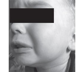
On the background of antibiotic therapy, the child had hyperthermic syndrome for 3 days. The body temperature returned to normal only for 4 following days. On the 7th day of treatment, the boy had a maculopapular rash on the skin of the abdomen, chest, without a tendency to merge. Single elements of the rash were noted on the extremities. On the 8th day of the treatment, facial rashes appeared (Fig. 1, 2). The tongue was covered with thick white coat. The bacterial flora of the throat and mouth was inoculated, and Str.viridans 103 CFU/ml, S.aureus 105 CFU/ml, Candida albicans 106 CFU/ml were detected in the throat. These pathogens are gene-rally recognized as the etiological factors of disease by conventional microbiological norms.
Evaluation of EBVI serological markers, abdominal ultrasound were performed to clarify the diagnosis.
Serum enzyme-linked immunoassay (BioPlex 2200 analyzer, TC BioRad (USA); EUROIMMUN (Germany)) confirmed the etiological factor of the underlying disease:
— EBV viral capsid antigen (VCA) IgM antibody index (AI) — 2.63 (more than 0.8 — a positive result);
— VCA IgG antibodies — 0.72 (below 0.8 — a negative result);
— measles virus, IgM AI — 0.56 (below 0.8 — a negative result);
— measles virus, IgG AI — 0.48 (below 0.8 — a negative result).
Abdominal ultrasound showed following in the liver: right lobe — 138 mm, middle-granular structure, usual echogenicity, left lobe — 69 mm. There was a torsion in the area of the gallbladder neck, the wall thickness — 2 mm. Pancreas: 20 × 13 × 22 mm, elevated echogenicity, middle-granular structure. The spleen was not enlarged.
Clinical diagnosis: Epstein-Barr viral infection, rhinopharyngitis, acute adenoiditis, acute otitis media. Mucocutaneous candidiasis of the oral cavity.

Discussion

We hypothesize that the similarity of clinical symptoms of infectious mononucleosis and oral candidiasis such as prolonged fever, lymphadenopathy, tonsillitis and exanthema syndrome complicates the diagnosis. The symptoms of oral candidiasis and mononucleosis are overlapped, and both diagnosis and treatment are complicated. We performed an extensive literature search in the Embase and Medline databases for articles published up to April 2020 to assess whether the search results were of relevance for the focus of this review. No language restrictions were applied. Systematic reviews, meta-analyses were prioritized. Our search results revealed that there are no published control studies of mixed Candida and EBVI. We found 54 articles on candidiasis. Of these, oral candidiasis has been analyzed in 20 papers. Clinical studies found a direct dependence between the prevalence of oral candidiasis and age of patients [4, 8, 12, 13]. We observed higher incidence of background somatic pathology, frequent use of antibiotics and hormones (steroid drugs) in case of candidiasis [13]. Disturbed reactions of immunoregulation and changes in resistance factors of a macroorganism, on one hand, and resistance of microorganisms to co-infection, on the other hand, determine the risk of development and severity of Candida lesion.
As shown by studies, primary EBVI is characterized by asymptomatic seroconversion of infection in 60–80 % of cases. In the remaining 20–40 % of those infected people, clinically manifested acute mononucleosis can develop. In the first two years of life, the proportion of latent forms reaches 90 %, in children aged 2–10 years, it decreases to 30–50 %. In infants, primary infection is more often characterized by an asymptomatic course than by the development of a typical pattern of infectious mononucleosis [4].
According to the literature findings, there may be several variants of EBVI effects, including clinical picture of secondary immunodeficiency, namely recurrent bacterial, fungal, often mixed infections of the respiratory and gastrointestinal tract [12]. White tonsillar exudate, sometimes even covering the tongue, may be seen, and they distinguish infectious mononucleosis from the more spotted coverings seen in bacterial tonsillitis [18].
Our literature review shows that mononucleosis infectiosa is characterized by fever, changes in the oropharynx, lymph nodes, liver, spleen, and peculiar changes in blood composition. Hematologic changes reminiscent of infectious mononucleosis can also occur with cytomegalovirus infection, toxoplasmosis, acute respiratory viral diseases, varicella, measles, infectious hepatitis, and other diseases [10]. Most of these heterophile antibody-negative cases of infectious mononucleosis-like infections are due to cytomegalovirus [6]. Classical triad of fever, sore throat, and generalized lymphadenopathy has been described in the literature [6, 15]. Hematologically, the disease is characterized by intensive lymphoproli-feration with atypical forms [5].
In our case, the patient had the following symptoms: white tongue, sore throat during swallowing, difficult nasal breathing, rashes on the skin of the trunk [14].
Our literature review shows that signs and symptoms of oral candidiasis may include the following: creamy white lesions on the tongue, inner cheeks, and sometimes on the roof of the mouth, gums, and tonsils; slightly increased lesions with a “cottage cheese”-like appearance; redness, burning or soreness that may be severe enough to cause difficulty with eating or swallowing; slight bleeding if the lesions are rubbed or scraped; cracking and redness at the corners of the mouth; cottony feeling in the mouth; loss of taste; redness, irritation, and pain under dentures (denture stomatitis) [11–13].
When conducting clinical diagnosis, one should pay attention to the common signs of candidiasis and mononucleosis, which make it difficult to carry out differential diagnosis at the clinical stage:
1. The appearance of rashes on the background or after receiving antibacterial therapy.
2. Persistent hyperthermia syndrome, which is difficult to control.
3. Presence of lymphadenopathy syndrome.
The diagnosis of mucocutaneous candidiasis or thrush usually can be made clinically. Yeast cells and pseudohyphae can be found in C.albicans-infected tissue and are detectable by microscopic examination of scrapings using Gram and calcofluor-white stain [35].
Infectious mononucleosis immunity and infection status can be assessed by:
1) the presence of at least classical clinical manifestations — fever, tonsillopharyngitis, cervical lymphadenopathy, hepatomegaly, or splenomegaly;
2) serological profile of primary EBVI — EBV VCA and Epstein-Barr virus nuclear antigen (EBNA) antibodies in the blood [7]. In these cases, in the acute phase, the test for VCA IgM antibodies is of diagnostic significance [6]. Additional testing for the presence of antibodies to EBNA may help differentiate acute and chronic infection. VCA and EBNA antibody tests have higher sensitivity and specificity. Few patients who have been infected with EBV will fail to develop antibodies to EBNA (approximately 5–10 %) [15, 16]. Articles focusing on EBV-associated infectious mononucleosis accentuate that the majority of infections can be recognized, however, by testing the patient’s serum for heterophile antibodies (rapid latex slide agglutination test; e.g., MONOS/Infectious Mononucleosis, Rapid Test, Serum), which usually appear within the first 3 weeks of illness, but then decline rapidly within a few weeks [5]. Heterophile antibody test is often unreliable in young children, particularly those under 4 years of age. Thus, assays specific for EBV must be performed in these cases, lest the diagnosis of infectious mononucleosis is missed [8]. EBV immunity and infection status can be assessed by testing EBV VCA and EBNA antibodies in blood [9]. Heterophile antibody test has high sensitivity and specificity and is considered the best initial tool for diagnosing infectious mononucleosis, but approximately 10 % of patients with this disease have a negative result.
We can summarize the key issues in the debate: if symptoms such as prolonged fever, white plaque on the tongue, exanthema on the mucous membrane, swollen lymph nodes, difficulty breathing through the nose, which are typical for both candidiasis and mononucleosis, occur, then it is necessary to perform differential diagnosis on C.albicans and exclude infectious mononucleosis. The interaction of C.albicans with the host innate immune system (caused by infectious mononucleosis) is the key factor in this immune imbalance. Despite a great number of studies on pediatric invasive candidiasis and EBVI, there is a lack of pediatric-specific recommendations as to examine the microbiota of the oropharynx in patients. Early diagnosis and, consequently, adequate treatment of invasive Candida infections is crucial to prevent mortality [16].

Conclusions

1. When infectious mononucleosis is combined with oral candidiasis, the following common symptoms occur: prolonged fever, lymphadenopathy, and exanthema syndrome. These symptoms usually occur after administration of broad-spectrum antibacterial agents.
2. In case of disease with symptoms such as prolonged fever, lymphadenopathy, tonsillitis, and exanthema syndrome, which are common for both oral candidiasis and infectious mononucleosis, differential diagnosis is required.
3. There is a need to examine the microbiota of the oropharynx in patients with infectious mononucleosis to detect flora, which may lead to the complicated course of the disease and diagnosis. In such case, inoculation of the oropharyngeal microbiota is an essential diagnostic tool for candidiasis detection.
Conflicts of interests. Authors declare the absence of any conflicts of interests and their own financial interest that might be construed to influence the results or interpretation of their manuscript.

Список литературы

  1. Voloha A.P. Epstein-Barr viral infection in children. Sovremennaya pediatriya. 2015. 4(68). 103-110 (in Ukrainian).
  2. Linke-Serinsöz E., Fend F., Quintanilla-Martinez L. Human immunodeficiency virus (HIV) and Epstein-Barr virus (EBV) related lymphomas, pathology view point. Semin. Diagn. Pathol. 2017. 34(4). 352-363.
  3. Chan C.W., Chiang A.K.S., Chan K.H. Epstein-Barr virus-associated infectious mononucleosis in Chinese children. Pediatric Infectious Disease Journal. 2003. 22(11). 974-978.
  4. Steinbach W.J., Roilides E., Berman D. Results from a prospective, international, epidemiologic study of invasive candidiasis in children and neonates. Pediatric Infectious Disease Journal. 2012. 31(12). 1252-1257.
  5. Manfredi R., Coronado O.V., Mastroianni A. Concurrent infectious mononucleosis and measles: a potentially life-threatening association sharing underlying immunodeficiency. Pediatric Infectious Disease Journal. 2003. 22(5). 470-471.
  6. Linde A., Falk K.I. Epstein-Barr virus. In: Manual of clinical microbiology. Ninth edition. Ed. by E.J. Barron, J.H. Jorgensen, M.L. Landry et al. ASM Press, 2007. 1564-1573.
  7. Setoh J.W.S., Ho C.K.M., Yung C.F. Epstein-Barr virus seroprevalence and force of infection in a multiethnic pediatric cohort, Singapore. Pediatric Infectious Disease Journal. 2019. 38(12). 1173-1176.
  8. Palazzi D.L., Arrieta A., Castagnola E. Candida speciation, antifungal treatment and adverse events in pediatric invasive candidiasis: results from 441 infections in a prospective, multi-national study. Pediatric Infectious Disease Journal. 2014. 33(12). 1294-1296.
  9. Pitetti R.D., Laus S., Wadowsky R.M. Clinical evaluation of a quantitative real time polymerase chain reaction assay for diagnosis of primary Epstein-Barr virus infection in children. Pediatric Infectious Disease Journal. 2003. 22(8). 736-739.
  10. Kramarev S.O., Vygovskaya O.V. Chronic forms of Epstein-Barr viral infection in children: current approaches to diagnosis and treatment. Sovremennaya pediatriya. 2008. 2(19). 103-107 (in Ukrainian).
  11. Martin J.M., Macias-Parra M., Mudry P. et al. Safety, efficacy, and exposure-response of voriconazole in pediatric patients with invasive aspergillosis, invasive candidiasis or esophageal candidiasis. Pediatric Infectious Disease Journal. 2017. 36(1). e1-e13.
  12. Lorenz M.C., Bender J.A., Fink G.R. Transcriptional response of Candida albicans upon internalization by macrophages. Eukaryotic Cell. 2004. 3(5). 1076-87.
  13. Autmizguine J., Tan S., Cohen-Wolkowiez M. et al. Antifungal susceptibility and clinical outcome in neonatal candidiasis. Pediatric Infectious Disease Journal. 2018. 37(9). 923-929.
  14. Fugl A., Andersen C.L. Epstein-Barr virus and its association with disease — a review of relevance to general practice. BMC Fam. Pract. 2019. 20. 62.
  15. Dunmire S.K., Hogquist K.A., Balfour H.H. Infectious mononucleosis. Curr. Top. Microbiol. Immunol. 2015. 390. 211-40.
  16. Sydnor E. Hospital epidemiology and infection control in acute-care settings. Clinical Microbiology Reviews. 2011. 24(1). 141-173.
  17. Azadmanesh J., Gowen A.M., Creger P.E., Schafer N.D., Blankenship J.R. Filamentation involves two overlapping, but distinct, programs of filamentation in the pathogenic fungus Candida albicans. G3: Genes, Genomes, Genetics. 2017. 7(11). 3797-3808.
  18. Grinstein S., Hube B., Mogavero S., Moran G., Westman J. Candida albicans hyphal expansion causes phagosomal membrane damage and luminal alkalinization. mBio. 2018. 9(5). e01226-18.
  19. Millsop J.W., Faze L.N. Oral candidiasis. Clin. Dermatol. 2016. 34. 487-494.
  20. Sherman R.G., Prusinski L., Joralmon R.A. Oral candidosis. Quintessence International. 2002. 33(7). 521-32.
  21. Vila T., Sultan A.S., Montelongo-Jauregui D., Jabra-Rizk M.A. Oral candidiasis: a disease of opportunity. Journal of Fungi. 2016. 6(1). 15.
  22. Kumamoto C.A. Candida biofilms. Current Opinion in Microbiology. 2002. 5(6). 608-11.
  23. Ortora G.J. Microbiology: an introduction. San Francisco, CA: Pearson Benjamin Cummings, 2010. 759.
  24. Peric M., Živkovic R., Milic Lemic A., Radunovic M., Milicic B., Arsic Arsenijevic V. The severity of denture stomatitis as related to risk factors and different Candida spp. Oral Surg. Oral Med. Oral Pathol. Oral Radiol. 2018. 126. 41-47.
  25. Singh R., Chakrabarti A. Invasive candidiasis in the Southeast-Asian region. In: Prasad R. (ed.). Candida albicans: cellular and molecular biology. 2nd ed. Switzerland: Springer International Publishing AG, 2017.
  26. Pfaller M.A., Diekema D.J. Epidemiology of invasive candidiasis: a persistent public health problem. Clinical Microbio-logy Reviews. 2007. 20(1). 133-63.
  27. Roilides E., Carlesse F. et al. Safety, efficacy and pharmacokinetics of anidulafungin in patients 1 month to < 2 years of age with invasive candidiasis, including candidemia. Pediatric Infectious Disease Journal. 2020. 39(4). 305-309.
  28. Tsekoura M., Ioannidou M., Pana Z.-D. Efficacy and safety of echinocandins for the treatment of invasive candidiasis in children: a meta-analysis. Pediatric Infectious Disease Journal. 2019. 38(1). 42-49.
  29. Benjamin D.K. Jr, Kaufman D.A., Hope W.W. A phase 3 study of micafungin versus amphotericin B deoxycholate in infants with invasive candidiasis. Pediatric Infectious Disease Journal. 2018. 37(10). 992-998.
  30. Kovanda L.L., Walsh T.J., Benjamin D.K. Jr. Exposure-response analysis of micafungin in neonatal candidiasis: pooled analysis of two clinical trials. Pediatric Infectious Disease Journal. 2018. 37(6). 580-585.
  31. Roilides E., Carlesse F., Leister-Tebbe H. A prospective, open-label study to assess the safety, tolerability and efficacy of anidulafungin in the treatment of invasive candidiasis in children 2 to < 18 years of age. Pediatric Infectious Disease Journal. 2019. 38(3). 275-279.
  32. Shkalim-Zemer V., Levi I., Fischer S. Response of symptomatic persistent chronic disseminated candidiasis to corticosteroid therapy in immunosuppressed pediatric patients: case study and review of the literature. Pediatric Infectious Disease Journal. 2018. 37(7). 686-690.
  33. Mason K.L., Erb Downward J.R., Mason K.D., Falkowski N.R., Eaton K.A., Kao J.Y., Young V.B., Hunagle G.B. Candida albicans and bacterial microbiota interactions in the cecum during recolonization following broad-spectrum antibiotic therapy. Infect. Immun. 2012. 80. 3371-3380.
  34. McIntyre G.T. Oral candidosis. Dent. Update. 2001. 28. 132-139.
  35. American Academy of Pediatrics, Committee on Infectious Diseases, Committee on Nutrition. Consumption of raw or unpasteurized milk and milk products by pregnant women and children. Pediatrics. 2014. 133(1). 175-179.

Вернуться к номеру