Архив офтальмологии Украины Том 9, №1, 2021
Вернуться к номеру
Етіологічні особливості бактеріального кератиту у хворих на цукровий діабет
Авторы: Заволока О.В.
Харківський національний медичний університет, м. Харків, Україна
Рубрики: Офтальмология
Разделы: Клинические исследования
Версия для печати
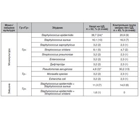
Мета роботи: вивчити етіологічні особливості бактеріального кератиту у хворих на цукровий діабет 1-го типу. Матеріали та методи. Аналіз здійснювався на основі даних обстеження 62 хворих на бактеріальний кератит та цукровий діабет 1-го типу та 43 хворих на бактеріальний кератит без цукрового діабету відповідного віку контрольної групи. Крім стандартних, методи офтальмологічного дослідження включали флюоресцеїновий тест, оптичну когерентну томографію переднього відрізку ока, безконтактне дослідження чутливості рогівки, бактеріологічне дослідження: посів культури з кон’юнктивальної порожнини на щільні поживні середовища (5% кров’яний агар та середовище для контролю стерильності), мікроскопічне дослідження мазка з кон’юнктиви із забарвленням за Романовським — Гімзою та Паппенгеймом. Результати. У хворих на цукровий діабет виявили особливості етіології бактеріального кератиту: грампозитивна мікрофлора була збудником у 1,5 раза частіше, а грамнегативна — у 3,1 раза рідше, ніж у хворих без цукрового діабету контрольної групи (р < 0,05). Крім того, Staphylococcus epidermidis був збудником бактеріального кератиту у хворих на цукровий діабет у 1,9 раза частіше, а Pseudomonas aeruginosa — у 6,3 раза рідше, ніж у хворих контрольної групи (р < 0,05). Висновки. У хворих на цукровий діабет відмічаються етіологічні особливості бактеріального кератиту, а саме переважання серед збудників грампозитивної мікрофлори за рахунок Staphylococcus epidermidis.
Background. The purpose was to study etiological features of bacterial keratitis in patients with type 1 diabetes mellitus. Materials and methods. The analysis was performed on the basis of survey data of 62 patients with bacterial keratitis and type 1 diabetes mellitus and 43 individuals with bacterial keratitis without diabetes mellitus of the corresponding age (control group). In addition to standard ones, ophthalmic methods included fluorescein test, anterior segment optical coherence tomography, non-contact corneal aesthesiometry, bacteriological examination: culture from the conjunctival cavity to dense nutrient media (5% blood agar and medium for sterility control), microscopic examination of conjunctival smear with Romanowsky-Giemsa and Pappenheim stain. Results. Etiological features of bacterial keratitis were found in patients with diabetes mellitus: Gram-positive microflora was the causative agent 1.5 times more often, and Gram-negative — 3.1 times less often than in patients without diabetes mellitus from the control group (p < 0.05). In addition, Staphylococcus epidermidis was the causative agent of bacterial keratitis in patients with diabetes mellitus 1.9 times more often, and Pseudomonas aeruginosa — 6.3 times less often than in patients of the control group (p < 0.05). Conclusions. There are etiological features of bacterial keratitis in patients with diabetes mellitus, namely predominance of the Gram-positive microflora among the pathogens due to Staphylococcus epidermidis.
цукровий діабет; бактеріальний кератит; етіологія
diabetes mellitus; bacterial keratitis; etiology
Вступ
Матеріали та методи
Результати та їх обговорення
Висновки
- Almizel A., Alsuhaibani F.A., Alkaff A.M., Alsaleh A.S., Al-Mansouri S.M. Bacterial Profile and Antibiotic Susceptibility Pattern of Bacterial Keratitis at a Tertiary Hospital in Riyadh. Clin. Ophthalmol. 2019, Dec 20. 13. 2547-2552. doi: 10.2147/OPTH.S223606. PMID: 31908410; PMCID: PMC6929922.
- Zagon I.S., Sassani J.W., Carroll M.A., McLaughlin P.J. Topical application of naltrexone facilitates reepithelialization of the cornea in diabetic rabbits. Brain Res. Bull. 2010. 81. 248-255.
- Badawi A.E., Moemen D., El-Tantawy N.L. Epidemiological, clinical and laboratory findings of infectious keratitis at Mansoura Ophthalmic Center, Egypt. Int. J. Ophthalmol. 2017, Jan 18. 10(1). 61-67. doi: 10.18240/ijo.2017.01.10. PMID: 28149778; PMCID: PMC5225350.
- Chang Y.S., Tai M.C., Ho C.H., Chu C.C., Wang J.J., Tseng S.H., Jan R.L. Risk of Corneal Ulcer in Patients with Diabetes Mellitus: A Retrospective Large-Scale Cohort Study. Sci. Rep. 2020, Apr 30. 10(1). 7388. doi: 10.1038/s41598-020-64489-0. PMID: 32355281; PMCID: PMC7193550.
- Wang B., Yang S., Zhai H.L., Zhang Y.Y., Cui C.X., Wang J.Y., Xie L.X. A comparative study of risk factors for corneal infection in diabetic and non-diabetic patients. Int. J. Ophthalmol. 2018, Jan 18. 11(1). 43-47. doi: 10.18240/ijo.2018.01.08. PMID: 29375989; PMCID: PMC5767656.
- Bilen H., Ates O., Astam N., Uslu H., Akcay G., Baykal O. Conjunctival flora in patients with type 1 or type 2 diabetes mellitus. Adv. Ther. 2007 Sep-Oct. 24(5). 1028-1035. doi: 10.1007/BF02877708. PMID: 18029329.
- Martins E.N., Alvarenga L.S., Höfling-Lima A.L., Freitas D., Zorat-Yu M.C., Farah M.E., Mannis M.J. Aerobic bacterial conjunctival flora in diabetic patients. Cornea. 2004 Mar. 23(2). 136-142. doi: 10.1097/00003226-200403000-00006. PMID: 15075882.
- Karimsab D., Razak S.K. Study of aerobic bacterial conjunctival flora in patients with diabetes mellitus. Nepal. J. Ophthalmol. 2013 Jan-Jun. 5(1). 28-32. doi: 10.3126/nepjoph.v5i1.7818. PMID: 23584643.
- Kruse A., Thomsen R.W., Hundborg H.H., Knudsen L.L., Sørensen H.T., Schønheyder H.C. Diabetes and risk of acute infectious conjunctivitis — a population-based case-control study. Diabet Med. 2006 Apr. 23(4). 393-397. doi: 10.1111/j.1464-5491.2006.01812.x. PMID: 16620267.
- Grzybowski A., Kanclerz P., Huerva V., Ascaso F.J., Tuuminen R. Diabetes and Phacoemulsification Cataract Surgery: Difficulties, Risks and Potential Complications. J. Clin. Med. 2019, May 20. 8(5). 716. doi: 10.3390/jcm8050716. PMID: 31137510; PMCID: PMC6572121.


/11.jpg)