Інформація призначена тільки для фахівців сфери охорони здоров'я, осіб,
які мають вищу або середню спеціальну медичну освіту.

Підтвердіть, що Ви є фахівцем у сфері охорони здоров'я.

Журнал "Гастроэнтерология" Том 55, №2, 2021

Вернуться к номеру

Раціональний підхід до профілактики тромбоемболічних ускладнень при хірургічному лікуванні ускладнених форм холелітіазу у хворих iз цирозом печінки

Авторы: S.D. Khimich(1), F.T. Muraviov(2)
(1) — National Pirogov Memorial Medical University, Vinnytsia, Ukraine
(2) — Zhytomyr Regional Clinical Hospital, Zhytomyr, Ukraine

Рубрики: Гастроэнтерология

Разделы: Клинические исследования

Версия для печати


Резюме

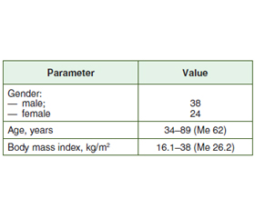
Актуальність. З літератури відомо, що значна частка ускладнень циррозу печінки пов’язана з негативним впливом цієї патології на систему згортання крові. За даними багатьох вчених, у більшості випадків при цирозі печінки існує ризик розвитку як тромботичних, так і геморагічних ускладнень. Мета дослідження: виявити раціональний підхід до профілактики тромбоемболічних ускладнень при хірургічному лікуванні ускладнених форм холелітіазу у хворих iз цирозом печінки. Матеріали та методи. Проведено ретроспективний аналіз історій хвороб 62 пацієнтів, які лікувалися з приводу ускладнених форм жовчнокам’яної хвороби та верифікованого цирозу печінки. Розподіл пацієнтів за нозологією був таким: гострий калькульозний холецистит — 48 осіб, синдром Міріззі — 7 та холедохолітіаз — 7. Результати. На основі порівняльної оцінки двох груп ризик інтраопераційної кровотечі в пацієнтів із передопераційною профілактикою був вищий (3 випадки — 8,1 %) при крововтраті понад 400 мл. Гематоми післяопераційних ран спостерігалися в 5 випадках у першій групі та в одному — у другій. У групі передопераційної профілактики розвинулися тромбоз ворітної вени (n = 1), тромбоз дрібних гілок легеневої артерії (n = 1) і тромбоз глибоких вен гомілки (n = 1). У групі післяопераційної профілактики таких ускладнень не спостерігалося. Висновки. Рішення щодо профілактики тромбоемболічних ускладнень у цієї категорії пацієнтів має бути зваженим і включати оцінку ризиків розвитку як геморагічних розладів, так і ускладнень, пов’язаних із тромбозом.

Background. It is known from scientific sources that a significant proportion of complications of liver cirrhosis is associated with negative impact of this pathology on the coagulation system. According to many scientists, liver cirrhosis in most cases poses a risk of developing both thrombotic and hemorrhagic complications. Objective: to identify a rational approach to the prevention of thromboembolic complications in the surgical treatment of complicated forms of cholelithiasis in patients with cirrhosis. Materials and methods. We retrospectively analyzed the hospital records of 62 patients who were treated for complicated forms of cholelithiasis with verified liver cirrhosis for the period from 2005 to 2018. The distribution of patients by nosology was as follows: acute calculous cholecystitis — 48 patients, Mirizzi syndrome — 7, choledocholithiasis — 7. Results. Based on a comparative analysis of two groups, the risk of intraoperative bleeding in patients with preoperative prophylaxis was higher (3 cases — 8.1 %), with a blood loss of more than 400 ml. Hematomas of postoperative wounds were observed in 5 cases in group 1 and in one case in group 2. In the group of preoperative prophylaxis, portal vein thrombosis (n = 1), thrombosis of small branches of the pulmonary artery (n = 1), and deep vein thrombosis of the leg (n = 1) developed. No such complications were observed in the group with postoperative prophylaxis. Conclusions. The decision on the prevention of thromboembolic complications in this category of patients should be balanced and include an assessment of the risks of developing both hemorrhagic disorders and complications associated with thrombosis.


Ключевые слова

жовчнокам’яна хвороба; цироз печінки; коагулопатії

gallstone disease; liver cirrhosis; coagulopathy

Introduction

Over the past decades, the development of minimally invasive surgical interventions has made it possible to significantly reduce the incidence of complications during the surgical treatment of complicated forms of cholelithiasis. Despite this, in most cases, the presence of concomitant liver cirrhosis leads to a significant increase in perioperative complications. According to many authors, the risk of postoperative complications in patients with concomitant liver cirrhosis can vary and reach 23–25 % [1–3]. A significant proportion of complications is associated precisely with the negative impact of cirrhosis on the coagulation system. According to the data of many scientists, liver cirrhosis in most cases poses a risk of developing both thrombotic and hemorrhagic complications. In the compensated stage of liver cirrhosis, the coagulation system is in a state of balance, which can be disturbed in the presence of an infectious agent and complicated forms of cholelithiasis. Despite the increased risk of hemorrhagic complications during surgical interventions against the background of liver cirrhosis, many authors note an increased risk of venous thromboembolism [4–8]. And yet, some scientists are not sure about the thromboprophylaxis for liver cirrhosis [9]. Thus, to date, there are no clear guidelines for the prevention of thromboembolic complications in the treatment of this category of patients.

Materials and methods

We retrospectively analyzed the hospital records of 62 patients who were treated for complicated forms of cholelithiasis with verified liver cirrhosis for the period from 2005 to 2018. In order to analyze and minimize the receipt of unreliable data, at the stage of planning the study, we have formed the criteria for selecting patients for this study. The selection criteria were as follows: the presence of complicated forms of cholelithiasis (acute calculous cholecystitis, choledocholithiasis and Mirizzi syndrome), mandatory surgery, and liver cirrhosis. Patients with a history of cancer and acute biliary pancreatitis were excluded from the study. In all cases, the class of liver cirrhosis was determined using the Child-Turcotte-Pugh score. Additionally, the assessment was carried out using the model for end-stage liver disease. The general condition of patients was assessed by means of the APACHE II score. In all cases, at the stage of diagnosis verification, standard laboratory and instrumental methods of examination were used, they necessarily included ultrasound examination of the abdominal organs, esophagofibrogastroduodenoscopy with mandatory assessment of the presence of varicose veins. Multislice computed tomography and magnetic resonance (MR) cholangiopancreatography were performed as indicated. Laboratory studies in all cases focused on the assessment of the coagulation system.
The distribution of patients depending on the class of cirrhosis:
— A — 34 (54.8 %);
— B — 26 (41.9 %);
— С — 2 (3.3 %).
The diagnosis of liver cirrhosis and the preoperative stage was verified in 51 (82.2 %) of 62 patients; in the rest of the cases, cirrhotic transformation of the liver was detected intraoperatively, and the diagnosis was finally established during liver biopsy.
The distribution of patients by age, sex and body mass index is presented in Table 1.
The distribution of patients by nosology was as follows: acute calculous cholecystitis — 48 cases, Mirizzi syndrome — 7, choledocholithiasis — 7. In the group of acute calculous cholecystitis, 2 patients had catarrhal form of gallbladder inflammation, 32 — acute phlegmonous cholecystitis, and phlegmonous-gangrenous changes were observed in 14 patients. In 23 cases, along with phlegmonous and phlegmonous-gangrenous forms, patients had complications such as gallbladder empyema (18), paravesical abscess (4), liver abscess (1). The diagnosis of Mirizzi syndrome was established at the preoperative stage in 6 of 7 patients. For disease staging, Сsendes and Beltran classification was used. Type I Mirizzi syndrome was diagnosed in 4 patients, type II — in 1, type III — in 1, type V — in 1 (the presence of a cholecystocolonic fistula). In 7 patients with a clinical picture of obstructive jaundice, calculi in the extrahepatic biliary tract were diagnosed using MR cholangiography. In one case, the diagnosis of choledocholithiasis was established during an ultrasound study, and MR cholangiography or endoscopic retrograde cholangiopancreatography was impossible due to the serious condition of the patient. These nosologies are compared with the class of liver cirrhosis in each case (Table 2).
At the stage of follow-up examination, all patients underwent mandatory standard instrumental examinations, which included electrocardiography, echocardiography, and chest X-ray. When choosing treatment and options for additional examination, a multidisciplinary approach was used with the involvement of surgeons, cardiologists, anesthesiologists, gastroenterologists and neuropathologists. Among the concomitant diseases identified, the prevailing ones were: chronic ischemic heart disease — 30, hypertension — 12, type 2 diabetes — 2, obesity — 5.
Based on the data of laboratory studies, coagulation disorders were detected in 45 patients (72.58 %). All patients underwent a comprehensive examination with an assessment of both coagulation and anticoagulation systems. The following indicators were analyzed without fail: fibrinogen level, prothrombin index, international normalized ratio, activated partial thromboplastin time, soluble fibrin monomer complexes, as well as the platelet hemostasis. D-dimer studies were carried out selectively (Table 3). 

Results and discussion

In all cases, since 2013, patients have been managed according to the Tokyo Guidelines. The main aspects of therapy were detoxification, antibacterial, hepatoprotective components. The nutritional component was prescribed by indications after determining the index according to G.P. Buzby or depending on the data of the point scale developed by the European Society for Medical Oncology. After conservative treatment, all patients underwent surgical interventions. The range of operations performed is presented in Table 4.
In all cases, we used a rational approach to the prevention of thromboembolic complications. Whenever possible, the strategy of fast track surgery was used in all patients in order to minimize the development of thromboembolic complications in the postoperative period. Prevention of thromboembolic complications was used in patients with a high and moderate risk of developing thromboembolic complications, with the exception of people at high risk of bleeding. Thus, the prevention of thromboembolic complications was carried out according to two schemes, the first of which was the standard preoperative administration of low molecular weight heparins (37 patients), and the second one included the early postoperative start of prophylaxis, 6 hours after completing the surgery (25 individuals). We have carried out a comparative analysis of 2 schemes for the surgical treatment of this category of patients. The main complications that arose as a result of coagulation disorders are presented in Table 5.
Based on the assessment of the two comparison groups, the risk of intraoperative bleeding in the group of patients with preoperative prophylaxis was higher (3 cases — 8.1 %), with a blood loss of more than 400 ml. Hematomas of postoperative wounds were detected in 5 cases in group 1 and in one case in group 2. In the group of preoperative prophylaxis, portal vein thrombosis (n = 1), thrombosis of small branches of the pulmonary artery (n = 1), and deep vein thrombosis of the leg developed. No such complications were observed in patients with postoperative prophylaxis.

Conclusions

Despite the small sample, it is possible to conclude that prophylaxis of thromboembolic complications in the early postoperative period can be used in selected categories of patients with a high risk of developing hemorrhagic complications. Fast track surgery strategy should be applied to every patient. The decision on the prevention of thromboembolic complications in this cohort should be balanced and include an assessment of the risks of developing both hemorrhagic disorders and complications associated with thrombosis.
 
Received 26.04.2021
Revised 12.05.2021
Accepted 19.05.2021

Список литературы

  1. Lledó J.B., Ibañez J.C., Mayor L.G. et al. Laparoscopic cholecystectomy and liver cirrhosis. Surg. Laparosc. Endosc. Percutan. Tech. 2011. 21. 391-5.
  2. Pinheiro R., Waisberg D., Lai Q., Andraus W., Nacif L., Rocha-Santos V., D’Albuquerque L. Laparoscopic cholecystectomy and cirrhosis: patient selection and technical considerations. Annals of Laparoscopic and Endoscopic Surgery. 2017. 2(3). doi: 10.21037/3784.
  3. Strömberg J., Hammarqvist F., Sadr-Azodi O., Sandblom G. Cholecystectomy in patients with liver cirrhosis. Gastroenterol. Res. Pract. 2015. 2015. 783823. doi: 10.1155/2015/783823.
  4. Abbas N., Makker J., Abbas H., Balar B. Perioperative care of patients with liver cirrhosis: a review. Health Services Insights. 2017 Jan. doi: 10.1177/1178632917691270.
  5. Buresi M., Hull R., Coffin C.S. Venous thromboembolism in cirrhosis: a review of the literature. Can. J. Gastroenterol. 2012. 26(12). 905-908. doi: 10.1155/2012/175849.
  6. García-Fuster M.J., Abdilla N., Fabiá M.J., Fernández C., Oliver V., Forner M.J. Enfermedad tromboembólica venosa y cirrosis hepática [Venous thromboembolism and liver cirrhosis]. Rev. Esp. Enferm. Dig. 2008 May. 100(5). 259-62. (in Spanish). doi: 10.4321/s1130-01082008000500002. PMID: 18662076.
  7. Khoury T., Ayman A.R., Cohen J., Daher S., Shmuel C., Mizrahi M. The complex role of anticoagulation in cirrhosis: an updated review of where we are and where we are going. Digestion. 2016. 93. 149-159. doi: 10.1159/000442877.
  8. O’Leary J.G., Greenberg C.S., Patton H.M., Caldwell S.H. AGA clinical practice update: coagulation in cirrhosis. Gastroenterology. 2019 Jul. 157(1). 34-43.e1. doi: 10.1053/j.gastro.2019.03.070. Epub 2019 Apr 12. PMID: 30986390.
  9. Senzolo M., Sartori M.T., Lisman T. Should we give thromboprophylaxis to patients with liver cirrhosis and coagulopathy? HPB (Oxford). 2009. 11(6). 459-464. doi: 10.1111/j.1477-2574.2009.00079.x.

Вернуться к номеру