Інформація призначена тільки для фахівців сфери охорони здоров'я, осіб,
які мають вищу або середню спеціальну медичну освіту.

Підтвердіть, що Ви є фахівцем у сфері охорони здоров'я.

Журнал «Травма» Том 24, №3, 2023

Вернуться к номеру

Значення заднього лігаментозного комплексу при травматичному ушкодженні ділянки грудопоперекового переходу Частина 1. Морфологія та біомеханіка

Авторы: Нехлопочин О.С., Вербов В.В., Чешук Є.В., Вороді М.В.
ДУ «Інститут нейрохірургії ім. акад. А.П. Ромоданова НАМН України», м. Київ, Україна

Рубрики: Травматология и ортопедия

Разделы: Справочник специалиста

Версия для печати


Резюме

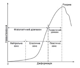
Відповідно до сучасних уявлень одним з базових критеріїв стабільності хребетно-рухового сегмента при травматичному його ушкодженні є цілісність заднього лігаментозного комплексу (ЗЛК). Щодо ділянки грудопоперекового переходу (ГПП) як зони, найвразливішої для травматичних ушкоджень, при визначенні тактики терапії використовують Thoracolumbar Injury Classification and Severity Score і клінічно орієнтовану класифікацію AOSpine Thoracolumbar Spine Injury Classification System, у якій стан ЗЛК є одним із трьох ключових параметрів. Уперше термін ЗЛК запропонував у 1963 р. F.W. Holdsworth. Однак лише широке впровадження в клінічну практику магнітно-резонансної томографії дало змогу повною мірою розкрити інформативність зазначеного параметра. До складу комплексу входять надостьова, міжостьова і жовта зв’язки і капсула фасеткових суглобів. Мета огляду: упорядкувати сучасні уявлення щодо морфології, біомеханічних особливостей, клінічної значущості й діагностичних можливостей виявлення ушкоджень ЗЛК при травматичних ушкодженнях ділянки ГПП. У першій частині детально розглянуто морфологічні й біомеханічні особливості ЗЛК зони ГПП. При описі морфології окремих зв’язок основну увагу приділено їхній протяжності, зонам фіксації та окремим шарам, якщо такі виділяють. Крім того, враховувалося взаємне анатомічне розташування структур, що розглядаються. Гістологічні особливості вказано лише в аспекті їхньої біомеханічної значущості. Аналіз даних літератури дав змогу упорядкувати дані, що характеризують біомеханічні параметри кожної зв’язки ЗЛК для кожного хребетно-рухового сегмента ділянки, що розглядається. Ураховували показники жорсткості, зусилля розриву, енергії руйнування, межі міцності й деформації, максимальної деформації та модуля пружності. Розглянуто особливості кривої «навантаження — деформація» зв’язкового апарату ділянки ГПП і методи розрахунку нелінійної зміни жорсткості кожної зв’язки ЗЛК у межах фізіологічних навантажень. Описано феномен переднавантаження і його клінічну значущість. Розглянуто деякі аспекти взаємодії елементів пасивної стабілізаційної системи при різних патернах навантаження. Наведені в першій частині огляду дані можуть бути корисними для загального розуміння принципів біомеханіки хребетно-рухового сегмента і можуть використовуватись при побудові високодеталізованих комп’ютерних моделей.

According to modern concepts, one of the basic criteria for the stability of the spinal motion segment in case of its traumatic damage is the integrity of the posterior ligamentous complex (PLC). Regarding the thoracolumbar junction (TLJ) as a zone that is most vulnerable to traumatic injuries, the Thoracolumbar Injury Classification and Severity Score and the clinically oriented AO Spine Thoracolumbar Spine Injury Classification System are used in determining therapeutic approaches in which the state of the thoracic spine is one of three key parameters. The term PLC was first proposed in 1963 by F.W. Holdsworth. However, only the widespread introduction of magnetic resonance imaging into clinical practice made it possible to fully reveal the informativeness of the specified parameter. The complex includes the interspinous, supraspinous ligaments, ligamentum flavum and facet joint capsule. The purpose of the review is to organize modern ideas about the morphology, biomechanical features, clinical significance, and diagnostic possibilities of detecting damage to the PLC in traumatic injuries of the TLJ area. In the first part, the morphological and biomechanical features of the PLC of the TLJ zone are considered in detail. When describing the morphology of some ligaments, the main attention is paid to their length, fixation zones, and certain layers, if such are distinguished. In addition, the relative anatomical location of the considered structures was taken into account. Histological features are indicated only in terms of their biomechanical significance. The analysis of literature data made it possible to organize the data characterizing the biomechanical parameters of each ligament of the PLC for each spinal motion segment of the area under consideration. Stiffness indicators, breaking force, fracture energy, strength and deformation limits, maximum deformation and elastic modulus were taken into account. The features of the load-deformation curve of the TLJ ligaments and methods of calculating the nonlinear change in the stiffness of each ligament of the PLC within the limits of physiological loads are considered. The phenomenon of preload and its clinical significance are described. Some aspects of the interaction between the elements of the passive stabilization system under different load patterns are considered. The data presented in the first part of the review can be useful for a general understanding of the principles of biomechanics of the spinal motion segment and may be used in the construction of highly detailed computer models.


Ключевые слова

задній лігаментозний комплекс; грудопоперековий перехід; хребетно-руховий сегмент; морфологія; біомеханіка

posterior ligamentous complex; thoracolumbar junction; spinal motion segment; morphology; biomechanics

Для ознакомления с полным содержанием статьи необходимо оформить подписку на журнал.


Список литературы

1. White A.A. 3rd, Johnson R.M., Panjabi M.M., Southwick W.O. Biomechanical analysis of clinical stability in the cervical spine. Clin. Orthop. Relat. Res. 1975. (109). 85-96. doi: 10.1097/00003086-197506000-00011. PMID: 1132209.
2. White A.A., Panjabi M.M. Clinical Biomechanics of the Spine. Lippincott, 1990.
3. Panjabi M.M., Kifune M., Liu W., Arand M., Vasavada A., Oxland T.R. Graded thoracolumbar spinal injuries: development of multidirectional instability. Eur. Spine J. 1998. 7(4). 332-339. doi: 10.1007/s005860050084. PMID: 9765043.
4. Vaccaro A.R., Lehman R.A. Jr., Hurlbert R.J., Anderson P.A., Harris M., Hedlund R. et al. A new classification of thoracolumbar injuries: the importance of injury morphology, the integrity of the posterior ligamentous complex, and neurologic status. Spine (Phila Pa 1976). 2005. 30(20). 2325-2333. doi: 10.1097/01.brs.0000182986.43345.cb. PMID: 16227897.
5. Vaccaro A.R., Oner C., Kepler C.K., Dvorak M., Schnake K., Bellabarba C. et al. AOSpine thoracolumbar spine injury classification system: fracture description, neurological status, and key modifiers. Spine (Phila Pa 1976). 2013. 38(23). 2028-2037. doi: 10.1097/BRS.0b013e3182a8a381. PMID: 23970107.
6. Bizdikian A.J., El Rachkidi R. Posterior Ligamentous Complex Injuries of the Thoracolumbar Spine: Importance and Surgical Implications. Cureus. 2021. 13(10). e18774. doi: 10.7759/cureus.18774. PMID: 34796064.
7. Holdsworth F. Fractures, dislocations and fracture-dislocations of the spine. J. Bone Joint Surg. Br. 1963. 45. 6-20.
8. Holdsworth F. Fractures, dislocations, and fracture-dislocations of the spine. J. Bone Joint Surg. Am. 1970. 52(8). 1534-1551. PMID: 5483077.
9. Louis R. Spinal stability as defined by the three-column spine concept. Anat. Clin. 1985. 7(1). 33-42. doi: 10.1007/BF01654627. PMID: 3994851.
10. Favero K.J., Van Peteghem P.K. The quadrangular fragment fracture. Roentgenographic features and treatment protocol. Clin. Orthop. Relat. Res. 1989. (239). 40-46. PMID: 2912635.
11. Magerl F., Aebi M., Gertzbein S.D., Harms J., Naza-rian S. A comprehensive classification of thoracic and lumbar injuries. Eur. Spine J. 1994. 3(4). 184-201. doi: 10.1007/BF02221591. PMID: 7866834.
12. Oner F.C., van Gils A.P., Dhert W.J., Verbout A.J. MRI findings of thoracolumbar spine fractures: a categorisation based on MRI examinations of 100 fractures. Skeletal Radiol. 1999. 28(8). 433-443. doi: 10.1007/s002560050542. PMID: 10486011.
13. Lee J.Y., Vaccaro A.R., Lim M.R., Oner F.C., Hulbert R.J., Hedlund R. et al. Thoracolumbar injury classification and severity score: a new paradigm for the treatment of thoracolumbar spine trauma. Journal of Orthopaedic Science: Official Journal of the Japanese Orthopaedic Association. 2005. 10(6). 671-675. doi: 10.1007/s00776-005-0956-y. PMID: 16307197.
14. Scapinelli R., Stecco C., Pozzuoli A., Porzionato A., Macchi V., De Caro R. The lumbar interspinous ligaments in humans: anatomical study and review of the literature. Cells, Tissues, Organs. 2006. 183(1). 1-11. doi: 10.1159/000094901. PMID: 16974090.
15. Neumann P., Wang Y., Kärrholm J., Malchau H., Nord-wall A. Determination of inter-spinous process distance in the lumbar spine. Evaluation of reference population to facilitate detection of severe trauma. Eur. Spine J. 1999. 8(4). 272-278. doi: 10.1007/s005860050172. PMID: 10483828.
16. Yahia L.H., Newman N., Rivard C.H. Neurohistology of lumbar spine ligaments. Acta Orthop. Scand. 1988. 59(5). 508-512. doi: 10.3109/17453678809148773. PMID: 2461043.
17. Iwanaga J., Simonds E., Yilmaz E., Schumacher M., Patel M., Tubbs R.S. Anatomical and Biomechanical Study of the Lumbar Interspinous Ligament. Asian Journal of Neurosurgery. 2019. 14(4). 1203-1206. doi: 10.4103/ajns.AJNS_87_19. PMID: 31903363.
18. Iida T., Abumi K., Kotani Y., Kaneda K. Effects of aging and spinal degeneration on mechanical properties of lumbar supraspinous and interspinous ligaments. Spine J. 2002. 2(2). 95-100. doi: 10.1016/s1529-9430(02)00142-0. PMID: 14588267.
19. Mori K., Yoshii T., Hirai T., Iwanami A., Takeuchi K., Yamada T. et al. Prevalence and distribution of ossification of the supra/interspinous ligaments in symptomatic patients with cervical ossification of the posterior longitudinal ligament of the spine: a CT-based multicenter cross-sectional study. BMC Musculoskelet Disord. 2016. 17(1). 492. doi: 10.1186/s12891-016-1350-y. PMID: 27903251.
20. Heylings D.J. Supraspinous and interspinous ligaments of the human lumbar spine. Journal of Anatomy. 1978. 125 (Pt 1). 127-131. PMID: 632208.
21. Chazal J., Tanguy A., Bourges M., Gaurel G., Escande G., Guillot M. et al. Biomechanical properties of spinal ligaments and a histological study of the supraspinal ligament in traction. J. Biomech. 1985. 18(3). 167-176. doi: 10.1016/0021-9290(85)90202-7. PMID: 3997901.
22. Viejo-Fuertes D., Liguoro D., Rivel J., Midy D., Guerin J. Morphologic and histologic study of the ligamentum flavum in the thoraco-lumbar region. Surgical and Radiologic Anatomy. 1998. 20(3). 171-176. doi: 10.1007/bf01628891. PMID: 9706675.
23. Chau A.M., Pelzer N.R., Hampton J., Smith A., Seex K.A., Stewart F. et al. Lateral extent and ventral laminar attachments of the lumbar ligamentum flavum: cadaveric study. Spine J. 2014. 14(10). 2467-2471. doi: 10.1016/j.spinee.2014.03.041. PMID: 24704501.
24. Olszewski A.D., Yaszemski M.J., White A.A. 3rd. The anatomy of the human lumbar ligamentum flavum. New observations and their surgical importance. Spine (Phila Pa 1976). 1996. 21(20). 2307-2312. doi: 10.1097/00007632-199610150-00001. PMID: 8915063.
25. Iwanaga J., Ishak B., Saga T., Singla A., Impastato D., Chapman J.R. et al. The Lumbar Ligamentum Flavum Does Not Have Two Layers and Is Confluent with the Interspinous Ligament: Anatomical Study with Application to Surgical and Interventional Pain Procedures. Clinical Anatomy (New York, NY). 2020. 33(1). 34-40. doi: 10.1002/ca.23437. PMID: 31325341.
26. Sato S., Oguma H., Murakami G., Noriyasu S. Morphometrical Study of the Joint Surface and Capsule of the Lumbar Zygapophysial Joint with Special Reference to Their Laterality. Okajimas Folia Anatomica Japonica. 2002. 79(1). 43-53. doi: 10.2535/ofaj.79.43.
27. Yamashita T., Minaki Y., Ozaktay A.C., Cavanaugh J.M., King A.I. A morphological study of the fibrous capsule of the human lumbar facet joint. Spine (Phila Pa 1976). 1996. 21(5). 538-543. doi: 10.1097/00007632-199603010-00002. PMID: 8852306.
28. Yahia L.H., Garzon S. Structure on the capsular ligaments of the facet joints. Annals of anatomy = Anatomischer Anzeiger: Official Organ of the Anatomische Gesellschaft. 1993. 175(2). 185-188. doi: 10.1016/s0940-9602(11)80179-2. PMID: 8489039.
29. Pintar F.A., Yoganandan N., Myers T., Elhagediab A., Sances A. Jr. Biomechanical properties of human lumbar spine ligaments. J. Biomech. 1992. 25(11). 1351-1356. doi: 10.1016/0021-9290(92)90290-h. PMID: 1400536.
30. Gardner-Morse M.G., Stokes I.A. Structural behavior of human lumbar spinal motion segments. J. Biomech. 2004. 37(2). 205-212. doi: 10.1016/j.jbiomech.2003.10.003. PMID: 14706323.
31. Dupuis P.R., Yong-Hing K., Cassidy J.D., Kirkaldy-Willis W.H. Radiologic diagnosis of degenerative lumbar spinal instability. Spine (Phila Pa 1976). 1985. 10(3). 262-276. doi: 10.1097/00007632-198504000-00015. PMID: 3992347.
32. Hukins D.W., Kirby M.C., Sikoryn T.A., Aspden R.M., Cox A.J. Comparison of structure, mechanical properties, and functions of lumbar spinal ligaments. Spine (Phila Pa 1976). 1990. 15(8). 787-795. PMID: 2237628.
33. Panjabi M.M., Goel V.K., Takata K. Physiologic strains in the lumbar spinal ligaments. An in vitro biomechanical study 1981 Volvo Award in Biomechanics. Spine (Phila Pa 1976). 1982. 7(3). 192-203. doi: 10.1097/00007632-198205000-00003. PMID: 6214027.
34. Allbrook D. Movements of the lumbar spinal column. J. Bone Joint Surg Br. 1957. 39-b(2). 339-345. doi: 10.1302/0301-620x.39b2.339. PMID: 13438978.
35. Von Forell G.A., Bowden A.E. Biomechanical implications of lumbar spinal ligament transection. Comput. Methods Biomech. Biomed. Engin. 2014. 17(15). 1685-1695. doi: 10.1080/10255842.2013.763936. PMID: 23477405.
36. Myklebust J.B., Pintar F., Yoganandan N., Cusick J.F., Maiman D., Myers T.J. et al. Tensile strength of spinal ligaments. Spine (Phila Pa 1976). 1988. 13(5). 526-531. PMID: 3187698.
37. Widmer J., Cornaz F., Scheibler G., Spirig J.M., Snedeker J.G., Farshad M. Biomechanical contribution of spinal structures to stability of the lumbar spine-novel biomechanical insights. Spine J. 2020. 20(10). 1705-1716. doi: 10.1016/j.spinee.2020.05.541. PMID: 32474224.
38. Panjabi M.M., Brand R.A. Jr, White A.A. 3rd. Three-dimensional flexibility and stiffness properties of the human thoracic spine. J. Biomech. 1976. 9(4). 185-192. doi: 10.1016/0021-9290(76)90003-8. PMID: 1262353.
39. Rohlmann A., Zander T., Schmidt H., Wilke H.J., Bergmann G. Analysis of the influence of disc degeneration on the mechanical behaviour of a lumbar motion segment using the finite element method. J. Biomech. 2006. 39(13). 2484-2490. doi: 10.1016/j.jbiomech.2005.07.026. PMID: 16198356.
40. Nachemson A.L., Evans J.H. Some mechanical pro-perties of the third human lumbar interlaminar ligament (ligamentum flavum). J. Biomech. 1968. 1(3). 211-220. doi: 10.1016/0021-9290(68)90006-7. PMID: 16329292.
41. Sengupta D.K., Herkowitz H.N. Degenerative spondylolisthesis: review of current trends and controversies. Spine (Phila Pa 1976). 2005. 30 (6 Suppl). S71-81. doi: 10.1097/01.brs.0000155579.88537.8e. PMID: 15767890.
42. Adams M.A., Hutton W.C., Stott J.R. The resistance to flexion of the lumbar intervertebral joint. Spine (Phila Pa 1976). 1980. 5(3). 245-253. doi: 10.1097/00007632-198005000-00007. PMID: 7394664.
43. Hindle R.J., Pearcy M.J., Cross A. Mechanical function of the human lumbar interspinous and supraspinous ligaments. Journal of Biomedical Engineering. 1990. 12(4). 340-344. doi: 10.1016/0141-5425(90)90010-k. PMID: 2395361.
44. Adams M.A., Hutton W.C. The relevance of torsion to the mechanical derangement of the lumbar spine. Spine (Phila Pa 1976). 1981. 6(3). 241-248. doi: 10.1097/00007632-198105000-00006. PMID: 7268544.
45. Meluzio M.C., Smakaj A., Perna A., Velluto C., Grillo G., Proietti L. et al. Epidemiology, diagnosis and management of Baastrup’s disease: a systematic review. J. Neurosurg. Sci. 2022. 66(6). 519-525. doi: 10.23736/s0390-5616.21.05428-x. PMID: 34342202.

Вернуться к номеру