Архив офтальмологии Украины Том 12, №2, 2024
Вернуться к номеру
Глікований гемоглобін як прогностичний фактор прогресування непроліферативної діабетичної ретинопатії при цукровому діабеті 2-го типу
Авторы: Сердюк А.В.
Дніпровський державний медичний університет, м. Дніпро, Україна
Рубрики: Офтальмология
Разделы: Клинические исследования
Версия для печати
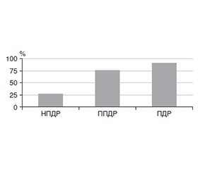
Актуальність. Перспективним напрямком сучасної медицини є підвищення точності прогнозу можливих результатів захворювання, його ускладнень або рецидивів. Для прогресування діабетичної ретинопатії (ДР) при цукровому діабеті (ЦД) 2-го типу мають значення декілька факторів, серед яких обговорюються глікований гемоглобін, тривалість ЦД 2-го типу та інші. Мета: визначити можливості прогнозу прогресування початкової непроліферативної ДР на підставі показників у крові глюкози, глікованого гемоглобіну та холестерину. Матеріали та методи. Обстежено 358 пацієнтів (358 очей) з ЦД 2-го типу та ДР, яких було розподілено на групи: 1-ша — з непроліферативною ДР (НПДР; 189 очей), 2-га — з препроліферативною ДР (ППДР, 96 очей) та 3-тя — з проліферативною ДР (ПДР; 73 ока). Пацієнти протягом 2 років були обстежені із застосуванням офтальмологічних методів, у сироватці крові визначали глюкозу натще, глікований гемоглобін та загальний холестерин колориметричним методом. Аналіз результатів дослідження проводився в пакеті EZR v.1.54 (Австрія). Результати. За віком та стажем ЦД 2-го типу суттєвої різниці між групами пацієнтів на початку дослідження виявлено не було, також і з прогресуванням ДР ці показники не мали зв’язку (р = 0,512 і р = 0,339 відповідно). Незалежними факторами ризику прогресування НПДР в однофакторному аналізі були вміст у крові глюкози (р = 0,002; ВШ = 1,08; 95% ВІ 1,03–1,13) та загального холестерину (р < 0,001; ВШ = 2,02; 95% ВІ 1,53–2,6). За вмістом у крові глікованого гемоглобіну була побудована логістична модель прогресування НПДР. Площа під кривою операційних характеристик AUC = 0,84 (95% ВІ 0,80–0,88), що свідчило про сильний зв’язок із прогресуванням НПДР. Поріг прогнозування вмісту глікованого гемоглобіну становив 8,9 % з чутливістю 75,6 % (95% ВІ 68,6–82 %) і специфічністю 79,9 % (95% ВІ 73,5–85,4 %). Висновки. Встановлено, що вміст у крові глікованого гемоглобіну, вищий за 8,9 %, є незалежним фактором прогресування НПДР, що дозволило побудувати прогностичну модель з дуже доброю якістю прогнозу.
Background. A promising direction of modern medicine is to increase the accuracy of predicting the possible outcomes of the disease, its complications or relapses. Several factors are important for the progression of diabetic retinopathy (DR) in type 2 diabetes (T2D), among which glycated hemoglobin, duration of T2D and others are discussed. The purpose was to determine the possibilities of predicting the progression of initial non-proliferative diabetic retinopathy (NPDR) based on the blood glucose, glycated hemoglobin and cholesterol indicators. Materials and methods. Three hundred and fifty-eight patients (358 eyes) with T2D and DR were examined and divided into groups: the first one — with NPDR (189 eyes), the second one — with pre-proliferative DR (96 eyes) and the third one — with proliferative DR (73 eyes). Patients were examined for 2 years using ophthalmological methods; serum fasting glucose, glycated hemoglobin and total cholesterol were determined by colorimetric method. The analysis of the research results was carried out in the EZR v. 1.54 package (Austria). Results. There was no significant difference between the groups at baseline in terms of age and T2D duration; these indicators were not associated with the DR progression (p = 0.512 and p = 0.339, respectively) as well. The independent risk factors for the NPDR progression in the univariate analysis were the content of blood glucose (p = 0.002; odds ratio (OR) = 1.08; 95% confidence interval (CI) 1.03–1.13) and total cholesterol (p < 0.001; OR = 2.02; 95% CI 1.53–2.6 %). Based on the glycated hemoglobin blood level, a logistic model of the NPDR progression was constructed. The area under the receiver operating characteristic curve was 0.84 (95% CI 0.80–0.88), which indicated a strong association with the NPDR progression. The threshold for predicting the glycated hemoglobin level was 8.9 % with a sensitivity of 75.6 % (95% CI 68.6–82 %) and a specificity of 79.9 % (95% CI 73.5–85.4 %). Conclusions. It was found that the content of glycated hemoglobin in the blood above 8.9 % is an independent factor for the NPDR progression, which allowed to build a prognostic model with a very good quality of prognosis.
непроліферативна діабетична ретинопатія; прогноз; прогресування; глікований гемоглобін; холестерин
non-proliferative diabetic retinopathy; prognosis; progression; glycated hemoglobin; cholesterol
Для ознакомления с полным содержанием статьи необходимо оформить подписку на журнал.
- Chung WK, Erion K, Florez JC, Hattersley AT, Hivert MF, Lee CG, et al. Precision medicine in diabetes: a Consensus Report from the American Diabetes Association (ADA) and the European Association for the Study of Diabetes (EASD). Diabetologia. 2020 Sep;63(9):1671-1693. doi: 10.1007/s00125-020-05181-w.
- Schiborn C, Schulze MB. Precision prognostics for the development of complications in diabetes. Diabetologia. 2022 Nov;65(11):1867-1882. doi: 10.1007/s00125-022-05731-4.
- Seid MA, Akalu Y, Gela YY, Belsti Y, Diress M, Fekadu SA, et al. Microvascular complications and its predictors among type 2 diabetes mellitus patients at Dessie town hospitals, Ethiopia. Diabetol Metab Syndr. 2021 Aug 17;13(1):86. doi: 10.1186/s13098-021-00704-w.
- Cardoso CRL, Leite NC, Dib E, Salles GF. Predictors of Development and Progression of Retinopathy in Patients with Type 2 Diabetes: Importance of Blood Pressure Parameters. Sci Rep. 2017 Jul 7;7(1):4867. doi: 10.1038/s41598-017-05159-6.
- Dagliati A, Marini S, Sacchi L, Cogni G, Teliti M, Tibollo V, et al. Machine Learning Methods to Predict Diabetes Complications. J Diabetes Sci Technol. 2018 Mar;12(2):295-302. doi: 10.1177/1932296817706375.
- Scanlon PH, Aldington SJ, Leal J, Luengo-Fernandez R, Oke J, Sivaprasad S, et al. Development of a cost-effectiveness model for optimisation of the screening interval in diabetic retinopathy screening. Health Technol Assess. 2015 Sep;19(74):1-116. doi: 10.3310/hta19740.
- Kanda Y. Investigation of the freely available easy-to-use software ‘EZR’ for medical statistics. Bone Marrow Transplant. 2013;48:452-8.
- Hintze JL, Nelson RD. Violin Plots: A Box Plot-Density Trace Synergism. The American Statistician. 1998;52(2):181-184. doi: 10.1080/00031305.1998.10480559.
- Schade DS, Shey L, Eaton RP. Cholesterol Review: A Metaboli–cally Important Molecule. Endocr Pract. 2020 Dec;26(12):1514-1523. doi: 10.4158/EP-2020-0347.
- Гур’янов В.Г., Лях Ю.Є., Парій В.Д., Короткий О.В., Чалий О.В., Чалий К.О. та ін. Посібник з біостатистики. Аналіз результатів медичних досліджень у пакеті EZR (R–statistics). Київ: Вістка, 2018. 208 с.
- Cho A, Park HC, Lee YK, Shin YJ, Bae SH, Kim H. Progression of Diabetic Retinopathy and Declining Renal Function in Patients with Type 2 Diabetes. J Diabetes Res. 2020 Jul 26;2020:8784139. doi: 10.1155/2020/8784139.
- Lee CC, Hsing SC, Lin YT, Lin C, Chen JT, Chen YH, et al. The Importance of Close Follow-Up in Patients with Early-Grade Diabetic Retinopathy: A Taiwan Population-Based Study Grading via Deep Learning Model. Int J Environ Res Public Health. 2021 Sep 16;18(18):9768. doi: 10.3390/ijerph18189768.
- Nordwall M, Abrahamsson M, Dhir M, Fredrikson M, Ludvigsson J, Arnqvist HJ. Impact of HbA1c, followed from onset of type 1 diabetes, on the development of severe retinopathy and nephropathy: the VISS Study (Vascular Diabetic Complications in Southeast Sweden). Diabetes Care. 2015 Feb;38(2):308-15. doi: 10.2337/dc14-1203.
- Jenkins AJ, Joglekar MV, Hardikar AA, Keech AC, OʼNeal DN, Januszewski AS. Biomarkers in diabetic retinopathy. Rev Diabet Stud. 2015 Spring-Summer;12(1-2):159-95. doi: 10.1900/RDS.2015.12.159.
- Testa R, Bonfigli AR, Prattichizzo F, La Sala L, De Nigris V, Ceriello A. The “Metabolic Memory” Theory and the early treatment of hyperglycemia in prevention of diabetic complications. Nutrients. 2017 Apr 28;9(5):437. doi: 10.3390/nu9050437.
- Perais J, Agarwal R, Evans JR, Loveman E, Colquitt JL, Owens D et al. Prognostic factors for the development and progression of proliferative diabetic retinopathy in people with diabetic retinopathy. Cochrane Database Syst Rev. 2023 Feb 22;2(2):CD013775. doi: 10.1002/14651858.CD013775.pub2.
- Gui YM, Guo J. The clinical significance of C-peptide for assessing the prognosis of non-proliferative diabetic retinopathy. Chinese Journal of Experimental Ophthalmology. 2013;31(8):775-8.
- Kim YJ, Kim JG, Lee JY, Lee KS, Joe SG, Park JY, et al. Development and progression of diabetic retinopathy and associated risk factors in Korean patients with type 2 diabetes: the experience of a tertiary center. J Korean Med Sci. 2014 Dec;29(12):1699-705. doi: 10.3346/jkms.2014.29.12.1699.
- Liu Y, Li J, Ma J, Tong N. The Threshold of the Severity of Diabetic Retinopathy below Which Intensive Glycemic Control Is Beneficial in Diabetic Patients: Estimation Using Data from Large Randomized Clinical Trials. J Diabetes Res. 2020 Jan 17;2020:8765139. doi: 10.1155/2020/8765139.
- Fullerton B, Jeitler K, Seitz M, Horvath K, Berghold A, Siebenhofer A. Intensive glucose control versus conventional glucose control for type 1 diabetes mellitus. Cochrane Database Syst Rev. 2014 Feb 14;2014(2):CD009122. doi: 10.1002/14651858.CD009122.pub2.
- Sivaprasad S, Pearce E. The unmet need for better risk strati–fication of non-proliferative diabetic retinopathy. Diabet Med. 2019 Apr;36(4):424-433. doi: 10.1111/dme.13868.
- Kataoka SY, Lois N, Kawano S, Kataoka Y, Inoue K, Watanabe N. Fenofibrate for diabetic retinopathy. Cochrane Database Syst Rev. 2023 Jun 13;6(6):CD013318. doi: 10.1002/14651858.CD013318.pub2.
- Srinivasan S, Raman R, Kulothungan V, Swaminathan G, Sharma T. Influence of serum lipids on the incidence and progression of diabetic retinopathy and macular oedema: Sankara Nethralaya Diabetic Retinopathy Epidemiology аnd Molecular genetics Study-II. Clin Exp Ophthalmol. 2017 Dec;45(9):894-900. doi: 10.1111/ceo.12990.

