Журнал "Гастроэнтерология" Том 58, №4, 2024
Вернуться к номеру
Фенотипи первинного склерозуючого холангіту в дітей
Авторы: Диба М.Б. (1), Березенко В.С. (1, 2)
(1) - ДУ «Всеукраїнський центр материнства та дитинства НАМН України», м. Київ, Україна
(2) - Національний медичний університет імені О.О. Богомольця, м. Київ, Україна
Рубрики: Гастроэнтерология
Разделы: Клинические исследования
Версия для печати
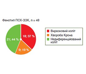
Актуальність. Первинний склерозуючий холангіт (ПСХ) у дітей — рідкісне хронічне імуноопосередковане захворювання жовчних шляхів, яке порівняно із захворюванням в дорослих має менш агресивний перебіг з ураженням внутрішньопечінкових жовчних проток і поєднується з автоімунним гепатитом, що створює особливий клінічний фенотип ПСХ — автоімунний склерозуючий холангіт (АСХ). Хоча імуносупресивна терапія ефективна для контролю автоімунного запалення, вона не гальмує прогресування фіброзних змін навколо жовчних проток, що, на жаль, призводить до формування біліарного цирозу печінки. Дослідження, спрямовані на вивчення клінічного перебігу ПСХ у дітей і вдосконалення ранньої діагностики, є актуальними, особливо з огляду на обмежений доступ до сучасних діагностичних методів, таких як магнітно-резонансна холангіопанкреатографія, потребу в інвазивних дослідженнях і відсутність стандартизованих діагностичних критеріїв, адаптованих до дитячого віку, що ускладнює діагностику і лікування цих пацієнтів. Мета дослідження: дослідити особливості клінічного перебігу первинного склерозуючого холангіту в дітей і підлітків залежно від фенотипу захворювання з метою розробки індивідуалізованих підходів до лікування. Матеріали та методи. Виконано ретроспективний і проспективний аналіз клінічного перебігу первинного склерозуючого холангіту в когорті дітей і підлітків, які лікувалися і спостерігалися у відділенні дитячої гепатології з 2016 по 2024 р. У дослідження включено 68 дітей (62 % хлопчиків і 38 % дівчаток) з ПСХ віком від 3 до 18 років (середній вік на момент встановлення діагнозу (11,0 ± 3,9) року), з яких у 38 (56 %) діагностовано автоімунний склерозуючий холангіт і в 30 (44 %) — ізольовану форму ПСХ (ІПСХ) без клінічних і гістологічних ознак автоімунного гепатиту. Результати. У 40 % дітей з ПСХ у дебюті захворювання мав місце фіброз печінки F3-F4 за METAVIR, з них у 24 % — цироз печінки (ЦП). Найбільш поширеним у дітей з ПСХ був змішаний фенотип з ураженням як великих, так і малих жовчних проток (63 %). ПСХ з ураженням тільки великої протоки мали 11 % дітей, з них у 70 % діагностовано ЦП, а ураження тільки малих жовчних проток діагностовано у 26 % дітей, з яких 12,5 % мали ЦП (р = 0,01). Запальне захворювання кишечника (ЗЗК) діагностовано у 84 % дітей з ПСХ, з яких 32 % мали виразковий коліт, 38 % — недиференційоване ЗЗК, 16 % — хворобу Крона. У 56 % мав місце панколіт, у 19 % дітей виявлено гістологічні ознаки ЗЗК за відсутності клінічних та ендоскопічних проявів. Безсимптомний перебіг ЗЗК мав місце в 58 % дітей. Особливістю клінічного перебігу АСХ на відміну від ІПСХ є вірогідно вища частота анемії (47 і 27 % відповідно, р = 0,03), висока концентрація IgG у сироватці крові (23,9 і 12,5 г/л відповідно, р < 0,01), вищі показники АЛТ, АСТ, загального білірубіну (всі р < 0,01), неінвазивних маркерів фіброзу (APRI, FIB-4, 2D-SWE) (р < 0,01; р = 0,01; р = 0,04 відповідно) в дебюті захворювання. У групі дітей з АСХ частіше діагностували фіброз печінки F3-F4 за METAVIR, ніж у групі з ІПСХ (73 і 40 % відповідно, р = 0,06). Ураження тільки великої жовчної протоки діагностовано лише в групі ІПСХ (р < 0,0009). За фенотипом ЗЗК діти з АСХ та ІПСХ не мали відмінностей. Висновки. ПСХ у дітей має два фенотипи захворювання (АСХ і ІПСХ), які зустрічаються майже з однаковою частотою. АСХ та ІПСХ мають схожі фенотипи ураження жовчних шляхів і кишечника, однак відрізняються за клінічним перебігом і підходами до лікування. Ураження тільки великої жовчної протоки в дітей з ПСХ пов’язане зі швидким формуванням фіброзу і цирозу печінки. Фенотип ПСХ з ураженням тільки малих жовчних проток має сприятливий перебіг у дітей. Більшість дітей із ПСХ мають фенотип ПСХ-ЗЗК. Активний діагностичний пошук уражень жовчних шляхів і кишечника в дітей з ПСХ сприятиме розробці ефективного персоналізованого підходу до лікування та моніторингу, що, у свою чергу, покращить прогноз захворювання.
Background. Primary sclerosing cholangitis (PSC) in children is a rare chronic immune-mediated disease of the biliary tract, which, unlike in adults, has a less aggressive course with damage to the intrahepatic bile ducts and is combined with autoimmune hepatitis, creating a special clinical phenotype of PSC, autoimmune sclerosing cholangitis (ASC). Although immunosuppressive therapy is effective in controlling autoimmune inflammation, it does not inhibit the progression of fibrotic changes around the bile ducts, which, unfortunately, leads to the formation of biliary cirrhosis of the liver. Research aimed at studying the clinical course of PSC in children and improving early diagnosis is relevant, especially given the limited access to modern diagnostic methods, such as magnetic resonance cholangiopancreatography, the need for invasive studies, and the lack of standardized diagnostic criteria adapted to childhood, which complicates the diagnosis and treatment of these patients. Objective: to investigate the clinical features of primary sclerosing cholangitis in children and adolescents depending on disease phenotype with the aim of developing individualized treatment approaches. Materials and methods. Retrospective and prospective analysis of the clinical course of primary sclerosing cholangitis in children and adolescents who were treated and followed at the Department of Pediatric Hepatology from 2016 to 2024 was conducted. The study included 68 children (62 % boys and 38 % girls) with PSC aged 3 to 18 years (mean age at diagnosis was (11.0 ± 3.9) years). Of these, 38 patients (56 %) had autoimmune sclerosing cholangitis, and 30 (44 %) had isolated PSC without clinical or histological signs of autoimmune hepatitis. Results. At disease onset, 40 % of children with PSC had liver fibrosis graded F3-F4 on METAVIR, with 24 % showing cirrhosis. The most common phenotype in children with PSC was a mixed one involving both large and small bile ducts (63 %). PSC with large bile duct involvement alone was observed in 11 % of cases, 70 % of these children were diagnosed with cirrhosis. Small duct involvement alone was present in 26 % of cases, with cirrhosis in 12.5 % (p = 0.01). Inflammatory bowel disease (IBD) was diagnosed in 84 % of children with PSC: 32 % had ulcerative colitis, 38 % had indeterminate IBD, and 16 % had Crohn’s disease. Pancolitis occurred in 56 % of cases, while 19 % of patients had histological signs of IBD without clinical or endoscopic manifestations. Asymptomatic IBD was diagnosed in 58 % of cases. The clinical course of ASC differed from isolated PSC with significantly higher rates of anemia (47 vs. 27 %, p = 0.03), elevated serum IgG levels (23.9 vs. 12.5 g/l, р < 0.01), and higher levels of alanine aminotransferase, aspartate aminotransferase, total bilirubin (p < 0.01 for all), non-invasive fibrosis markers (APRI, FIB-4, 2D-SWE) at onset (p < 0.01; p = 0.01; p = 0.04, respectively). Liver fibrosis F3-F4 on METAVIR was more frequently diagnosed in ASC group than in patients with isolated PSC (73 vs. 40 %, p = 0.06). Large bile duct involvement alone was found only in isolated PSC (p < 0.0009). No differences were observed between ASC and isolated PSC in terms of IBD phenotype. Conclusions. PSC in children is represented by 2 phenotypes (ASC and isolated PSC) that occur with almost equal frequency. ASC and isolated PSC have similar phenotypes of biliary and intestinal lesions, however, they differ in terms of clinical course and therapeutic approaches. Involvement of the large bile duct alone in children with PSC is associated with rapid formation of liver fibrosis and cirrhosis. PSC phenotype with involvement of small bile ducts alone has a favorable course in children. Most children with PSC exhibit the PSC-IBD phenotype. Active diagnostic search for biliary and intestinal lesions in children with PSC will facilitate the development of effective personalized approaches to treatment and monitoring, thereby improving disease prognosis.
первинний склерозуючий холангіт; автоімунний склерозуючий холангіт; запальне захворювання кишечника; діти
primary sclerosing cholangitis; autoimmune sclerosing cholangitis; inflammatory bowel disease; children
Для ознакомления с полным содержанием статьи необходимо оформить подписку на журнал.
- Deneau M et al. Primary sclerosing cholangitis, autoimmune hepatitis, and overlap in Utah children: Epidemiology and natural history. Hepatology. 2013;58:1392-1400. DOI: 10.1002/hep.26454.
- Stevens JP, Gupta NA. Recent Insights into Pediatric Primary Sclerosing Cholangitis. Clin Liver Dis. 2022;26:489-519. DOI: 10.1016/j.cld.2022.03.009.
- Karlsen TH, Hirschfield GM. Primary Sclerosing Cholangitis, Current Understanding, Management, and Future Developments. In: Comprehensive Clinical Hepatology. 2016;99-110. DOI: 10.1007/978-3-319-40908-5_8.
- Deneau MR et al. The natural history of primary sclerosing cholangitis in 781 children: A multicenter, international collaboration. Deneau Hepatology. 2017;66:518-527. DOI: 10.1002/hep.29204.
- Valentino PL et al. The Natural History of Primary Sclerosing Cholangitis in Children. J Pediatr Gastroenterol Nutr. 2016;63:603-609. DOI: 10.1097/mpg.0000000000001368.
- Beretta-Piccoli BT, Vergani D, Mieli-Vergani G. Autoimmune sclerosing cholangitis: Evidence and open questions. J Autoimmun. 2018;95:15-25. doi: 10.1016/j.jaut.2018.10.008.
- Ricciuto A, Kamath BM, Hirschfield GM, Trivedi PJ. Primary sclerosing cholangitis and overlap features of autoimmune hepatitis: A coming of age or an age-ist problem? J Hepatol. 2023;79:567-575. DOI: 10.1016/j.jhep.2023.02.030.
- Mieli-Vergani G et al. Diagnosis and Management of Pediatric Autoimmune Liver Disease. J Pediatr Gastroenterol Nutr. 2018;66:345-360. DOI: 10.1097/mpg.0000000000001801.
- Gregorio GV et al. Autoimmune hepatitis/sclerosing cholangitis overlap syndrome in childhood: A 16-year prospective study. Hepato–logy. 2001;33:544-553. DOI: 10.1053/jhep.2001.22131.
- Deneau MR. The Sclerosing Cholangitis Outcomes in Pediat–rics (SCOPE) Index: A Prognostic Tool for Children. Hepatology. 2021;73:1074-1087. DOI: 10.1002/hep.31393.
- Munster KN van, Bergquist A, Ponsioen CY. Inflammatory bowel disease and primary sclerosing cholangitis: One disease or two? J Hepatol. 2024;80:155-168. DOI: 10.1016/j.jhep.2023.09.031.
- Kim YS, Hurley EH, Park Y, Ko S. Primary sclerosing cholangitis (PSC) and inflammatory bowel disease (IBD): a condition exemplifying the crosstalk of the gut-liver axis. Exp Mol Med. 2023;55:1380-1387. DOI: 10.1038/s12276-023-01042-9.
- Catassi G et al. Outcome of Very Early Onset Inflammatory Bowel Disease Associated With Primary Sclerosing Cholangitis: A Multicenter Study From the Pediatric IBD Porto Group of ESPGHAN. Inflamm Bowel Dis. 2023;30:1662-1669. DOI: 10.1093/ibd/izad218.
- Björnsson E. Small-duct primary sclerosing cholangitis. Curr Gastroenterol Rep. 2009;11:37-41. DOI: 10.1007/s11894-009-0006-6.
- Assis DN, Bowlus CL. Recent Advances in the Management of Primary Sclerosing Cholangitis. Clin Gastroenterol Hepatol. 2023;21:2065-2075. DOI: 10.1016/j.cgh.2023.04.004.
- Hensel KO et al. Sclerosing Cholangitis in Pediatric Inflammatory Bowel Disease: Early Diagnosis and Management Affect Clinical Outcome. J Pediatr. 2021;238:50-56.e3. doi: 10.1016/j.jpeds.2021.07.047.
- Faubion WA Jr, Loftus EV, Sandborn WJ, Freese DK, Perrault J. Pediatric “PSC-IBD”: A Descriptive Report of Associated Inflammatory Bowel Disease Among Pediatric Patients With PSC. J Pediatr Gastroenterol Nutr. 2001;33:296-300. DOI: 10.1097/00005176-200109000-00009.
- Ricciuto А. et al. Oral vancomycin is associated with improved inflammatory bowel disease clinical outcomes in primary sclerosing cholangitis-associated inflammatory bowel disease (PSC-IBD): A matched analysis from the Paediatric PSC Consortium. Aliment Pharmacol Ther. 2024;59:1236-1247. DOI: 10.1111/apt.17936.
- Ali AH et al. Open-label prospective therapeutic clinical trials: oral vancomycin in children and adults with primary sclerosing cholangitis. Scand J Gastroenterol. 2020;55:941-950. DOI: 10.1080/00365521.2020.1787501.
- Shah А. et al. Effects of Antibiotic Therapy in Primary Sclero–sing Cholangitis with and without Inflammatory Bowel Disease: A Systematic Review and Meta-Analysis. Semin Liver Dis. 2019;39:432-441. DOI: 10.1055/s-0039-1688501.
- Kim YS, Hurley EH, Park Y, Ko S. Treatment of primary sclerosing cholangitis combined with inflammatory bowel disease. Intest Res. 2023;21:420-432. DOI: 10.5217/ir.2023.00039.
- Trivedi PJ et al. Effects of Primary Sclerosing Cholangitis on Risks of Cancer and Death in People With Inflammatory Bowel Di–sease, Based on Sex, Race, and Age. Gastroenterology. 2020;159:915-928. DOI: 10.1053/j.gastro.2020.05.049.
- Bowlus CL et al. AASLD practice guidance on primary sclerosing cholangitis and cholangiocarcinoma. Hepatology. 2023;77:659-702. DOI: 10.1002/hep.32771.

