Журнал "Гастроэнтерология" Том 58, №4, 2024
Вернуться к номеру
Сироваткові біомаркери в діагностиці фіброзу печінки в дітей із метаболічно-асоційованою стеатотичною хворобою печінки
Авторы: Yu.M. Stepanov, N.Yu. Zavhorodnia, I.A. Klenina, O.M. Tatarchuk, O.P. Petishko
State Institution “Institute of Gastroenterology of the National Academy of Medical Sciences of Ukraine”, Dnipro, Ukraine
Рубрики: Гастроэнтерология
Разделы: Клинические исследования
Версия для печати
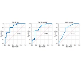
Актуальність. Епідемія COVID-19 та війна в Україні призвели до значного зростання кількості дітей, які страждають на метаболічно-асоційовану стеатотичну хворобу печінки (МАСХП). Однією з невирішених проблем, пов’язаних із МАСХП, лишається ідентифікація осіб із ризиком швидкого прогресування хвороби й розвитку необоротних змін печінки. Пошук альтернативних неінвазивних маркерів, придатних для раннього виявлення фіброзу печінки в дітей, надзвичайно актуальний. Мета: визначити діагностичну цінність сироваткових маркерів фіброзу та їхній зв’язок із сонографічними показниками й параметрами складу тіла в дітей із МАСХП. Матеріали та методи. У дослідження «випадок — контроль» включено 80 дітей віком від 6 до 17 років (у середньому (12,15 ± 2,51) року). Наявність стеатозу й фіброзу печінки визначали шляхом транзієнтної еластографії (Fibroscan® 502 touch F60156, Echosens, Франція). Усім обстеженим проведені антропометричні дослідження з оцінкою індексу маси тіла. При значенні останнього в межах 1–2 Z-score діагностували надмірну вагу, при перевищенні двох Z-score — ожиріння. За даними транзієнтної еластографії та індексу маси тіла всі діти були розділені на чотири групи: І — 27 дітей із МАСХП та фіброзом ≥ F1, ІІ — 35 дітей із МАСХП без фіброзу, ІІІ — 18 дітей із ожирінням або надмірною вагою без МАСХП і фіброзу. Контрольну ІV групу становили 14 дітей із нормальною вагою без МАСХП та фіброзу. Групи не мали значущих відмінностей за віковим і статевим розподілом. Дослідження складу тіла проведено шляхом біоімпедансометрії за допомогою аналізатора TANITA MC-780MA (виробник Maeno-cho, Itabashi-ku, Токіо, Японія). Концентрацію ендотеліального фактора росту судин (ЕФРС) в сироватці крові визначали за допомогою імуноферментного аналізу (ІФА) з використанням тест-систем фірми Wuhan Fine Biotech Co., Ltd (КНР) відповідно до рекомендацій виробника. Рівень цитокератину-18 (ЦК-18) у сироватці крові оцінювали за допомогою наборів IDL Biotech AB (Швеція) для ІФА. Уміст трансформуючого фактора росту бета-1 (ТФР-β1) визначали із застосуванням тест-системи ІФА IBL International (ФРН). Процеси фіброгенезу оцінювали за вмістом у сироватці крові гідроксипроліну вільного (ГПв), гідроксипроліну білковозв’язаного (ГПб/зв) та глікозаміногліканів (ГАГ). Результати. Продемонстровано вірогідне зростання рівнів ЦК-18 і ТФР-β1 у дітей із МАСХП-асоційованим фіброзом печінки. В осіб із фіброзом печінки спостерігалося підвищення співвідношення ГПв/ГПб/зв і рівня ГАГ у сироватці крові порівняно з пацієнтами з МАСХП без фіброзу й дітьми з надмірною вагою та ожирінням. Пороговий рівень ЦК-18 для діагностики фіброзу печінки в дітей із МАСХП становив 90,3 Од/л (чутливість 81,3 %, специфічність 76,9 %, AUC 0,843; р < 0,001). Чутливість порогового значення сироваткового рівня ТФР-β1 (96,8 пг/мл) у пацієнтів із МАСХП для діагностики фіброзу печінки дорівнювала 80,0 %, специфічність — 65,7 %, AUC — 0,787 (р < 0,001). Пороговий рівень ГАГ у сироватці крові понад 4,24 ммоль/л демонструє чутливість 70,6 % і специфічність 69,6 %, AUC 0,743 (р < 0,01). Продемонстровано позитивну кореляцію ЦК-18, ТФР-β1, ГАГ із показниками жорсткості й еластичності печінки, компонентним складом тіла дітей із МАСХП та високі рівні діагностичної точності, що дозволяє використовувати їх для скринінгу МАСХП-асоційованого фіброзу печінки в дітей. Висновки. Для дітей із фіброзом печінки характерний підвищений вміст ЦК-18, ЕФРС, ТФР-β1, ГПб/зв та ГАГ у сироватці крові. Порогові значення ЦК-18 (більше 90,3 Од/л), ТФР-β1 (понад 96,8 пг/мл) та ГАГ (більше 4,24 ммоль/л) мають високі показники чутливості й специфічності, що дозволяє використовувати їх для діагностики фіброзу печінки в дітей із МАСХП.
Background. The COVID-19 epidemic and the war in Ukraine have led to a significant increase in the number of children suffering from metabolic dysfunction-associated steatotic liver disease (MASLD). One of the unresolved problems associated with MASLD is the identification of individuals at risk of rapid disease progression and development of irreversible liver changes. The search for alternative noninvasive markers suitable for the early detection of liver fibrosis in children remains extremely relevant. The aim of the study was to determine the diagnostic value of serum fibrosis markers and their relationship with sonographic and body composition parameters in children with MASLD. Materials and methods. The case-control study included 80 children aged 6 to 17 years (mean of (12.15 ± 2.51) years). The presence of steatosis and liver fibrosis was determined by transient elastography (FibroScan® 502 touch F60156, Echosens, France). All subjects underwent anthropometric studies to determine body mass index. If it was within one-two Z-score, overweight was diagnosed. If the body mass index exceeded two Z-score, obesity was diagnosed. According to transient elastography and body mass index, all children were divided into four groups: group I — 27 children with MASLD and fibrosis ≥ F1, group II — 35 children with MASLD without fibrosis, group III — 18 obese or overweight children without MASLD and without fibrosis. The control group IV consisted of 14 children with normal weight without MASLD and without fibrosis. The groups had no significant differences in age and gender distribution. The study of body composition was performed by bioimpedance analysis using a TANITA MC-780MA analyzer (manufactured by Maeno-cho, Itabashi-ku, Tokyo, Japan). Quantitative determination of the serum concentration of vascular endothelial growth factor (VEGF) was performed by enzyme-linked immunosorbent assay (ELISA) using test systems from Wuhan Fine Biotech Co., Ltd (China) according to the manufacturer’s recommendations. The level of serum cytokeratin 18 (CK-18) was evaluated with IDL Biotech AB kits (Sweden) for ELISA. Serum content of transforming growth factor beta 1 (TGF-β1) was studied using an ELISA test system from IBL International (Germany). Fibrogenesis processes were evaluated by the serum content of free hydroxyproline (HPf), protein-bound hydroxyproline (HPp/b) and glycosaminoglycans (GAG). Results. The study revealed a significant increase in the level of CK-18 and TGF-β1 in children with MASLD-associated liver fibrosis. In children with liver fibrosis, an increase in the ratio of HPf/HPp/b and the level of GAG in the blood serum was observed compared to patients with MASLD without fibrosis and with overweight and obese children. The threshold value of CK-18 for liver fibrosis diagnosis was 90.3 U/l (sensitivity 81.3 %, specificity 76.9 %, AUC 0.843, p < 0.001). The sensitivity of the threshold value of serum TGF-β1 (96.8 pg/mL) in children with MASLD was 80.0 %, specificity 65.7 %, AUC 0.787 (p < 0.001). Threshold value of serum GAG (4.24 mmol/L) demonstrated a sensitivity of 70.6 % and a specificity of 69.6 %, AUC 0.743 (p < 0.01). CK-18, TGF-β1, GAG shown a positive correlation with liver stiffness and elasticity, body composition of MASLD children and had high levels of diagnostic accuracy, which allows them to be used in children when screening for MASLD-associated liver fibrosis. Conclusions. Children with liver fibrosis are characterized by elevated serum levels of CK-18, VEGF, TGF-β1, HPp/b and GAG. The threshold values of CK-18 (more than 90.3 U/l), TGF-β1 (above 96.8 pg/mL) and GAG (more than 4.24 mmol/l) have high sensitivity and specificity, which allows them to be used for the diagnosis of liver fibrosis in children with MASLD.
діти; метаболічно-асоційована стеатотична хвороба печінки; фіброз; цитокератин-18; глікозаміноглікани; трансформуючий фактор росту бета-1
children; metabolic dysfunction-associated steatotic liver disease; fibrosis; cytokeratin 18; glycosaminoglycans; transforming growth factor beta 1
Для ознакомления с полным содержанием статьи необходимо оформить подписку на журнал.
- Furthner D, Weghuber D, Dalus C, et al. Nonalcoholic Fatty Liver Disease in Children with Obesity: Narrative Review and Research Gaps. Hormone research in paediatrics. 2022;95(2):167-176. doi: 10.1159/000518595.
- Zelber-Sagi S, Carrieri P, Pericàs JM, Ivancovsky-Wajcman D, Younossi ZM, Lazarus JV. Food inequity and insecurity and MASLD: burden, challenges, and interventions. Nat Rev Gastroenterol Hepatol. 2024 Oct;21(10):668-686. doi: 10.1038/s41575-024-00959-4.
- Rupasinghe K, Hind J, Hegarty R. Updates in Metabolic Dysfunction-Associated Fatty Liver Disease (MAFLD) in Children. Journal of pediatric gastroenterology and nutrition. 2023;77(5):583-591. doi: 10.1097/MPG.0000000000003919.
- Hydes T, Brown E, Hamid A, Bateman AC, Cuthbertson DJ. Current and Emerging Biomarkers and Imaging Modalities for Nonalcoholic Fatty Liver Disease: Clinical and Research Applications. Clinical therapeutics. 2021;43(9):1505-1522. doi: 10.1016/j.clinthera.2021.07.012.
- Simon TG, Roelstraete B, Hartjes K, Shah U, Khalili H, Arnell H, Ludvigsson JF. Non-alcoholic fatty liver disease in children and young adults is associated with increased long-term mortality. J Hepatol. 2021 Nov;75(5):1034-1041. doi: 10.1016/j.jhep.2021.06.034.
- Zoncapè M, Liguori A, Tsochatzis EA. Non-invasive testing and risk-stratification in patients with MASLD. Eur J Intern Med. 2024 Apr;122:11-19. doi: 10.1016/j.ejim.2024.01.013.
- Draijer L, Voorhoeve M, Troelstra M, et al. A natural history study of paediatric non-alcoholic fatty liver disease over 10 years. JHEP Rep. 2023 Jan 25;5(5):100685. doi: 10.1016/j.jhepr.2023.100685.
- Vos MB, Abrams SH, Barlow SE, et al. NASPGHAN Clinical Practice Guideline for the Diagnosis and Treatment of Nonalcoholic Fatty Liver Disease in Children: Recommendations from the Expert Committee on NAFLD (ECON) and the North American Society of Pediatric Gastroenterology, Hepatology and Nutrition (NASPGHAN). J Pediatr Gastroenterol Nutr. 2017 Feb;64(2):319-334. doi: 10.1097/MPG.0000000000001482.
- Kalveram L, Baumann U, De Bruyne R, et al.; ESPGHAN Fatty Liver Special Interest Group. Noninvasive scores are poorly predictive of histological fibrosis in paediatric fatty liver disease. J Pediatr Gastroenterol Nutr. 2024 Jan;78(1):27-35. doi: 10.1002/jpn3.12068.
- Dezsőfi A, Baumann U, Dhawan, et al.; ESPGHAN Hepatology Committee. Liver biopsy in children: position paper of the ESPGHAN Hepatology Committee. J Pediatr Gastroenterol Nutr. 2015 Mar;60(3):408-20. doi: 10.1097/MPG.0000000000000632.
- Chen BR, Pan CQ. Non-invasive assessment of fibrosis and steatosis in pediatric non-alcoholic fatty liver disease. Clin Res Hepatol Gastroenterol. 2022 Jan;46(1):101755. doi: 10.1016/j.clinre.2021.101755.
- Draijer LG, van Oosterhout JPM, Vali Y, et al. Diagnostic accuracy of fibrosis tests in children with non-alcoholic fatty liver di–sease: A systematic review. Liver Int. 2021 Sep;41(9):2087-2100. doi: 10.1111/liv.14908.
- Younossi ZM, Loomba R, Anstee QM, et al. Diagnostic modalities for nonalcoholic fatty liver disease, nonalcoholic steatohepatitis, and associated fibrosis. Hepatology. 2018 Jul;68(1):349-360. doi: 10.1002/hep.29721.
- Horn P, Tacke F. Metabolic reprogramming in liver fibrosis. Cell Metab. 2024 Jul 2;36(7):1439-1455. doi: 10.1016/j.cmet.2024.05.003.
- Ullah A, Singla RK, Batool Z, Cao D, Shen B. Pro- and anti-inflammatory cytokines are the game-changers in childhood obesity-associated metabolic disorders (diabetes and non-alcoholic fatty liver diseases). Rev Endocr Metab Disord. 2024 Aug;25(4):783-803. doi: 10.1007/s11154-024-09884-y.
- Shalby MM, Ibrahim SA, Behairy OG, Behiry EG, Mahmoud DA. Diagnostic value of serum cytokeratin-18 in children with chronic liver disease. J Paediatr Child Health. 2020 Jan;56(1):41-46. doi: 10.1111/jpc.14488.
- Hegmar H, Wiggers T, Nasr P, et al. Performance of novel collagen turnover biomarkers to detect increased liver stiffness in MASLD. J Intern Med. 2024 Aug;296(2):177-186. doi: 10.1111/joim.13813.
- Grønbæk H, Mellemkjær A, Nielsen S, Magkos F. The vascular endothelial growth factor system — a new player in the pathogenesis and development of metabolic dysfunction-associated steatotic liver disease. Hepatobiliary Surg Nutr. 2023 Dec 1;12(6):963-965. doi: 10.21037/hbsn-23-552.
- Vali Y, Lee J, Boursier J, et al. Liver Investigation: Testing Marker Utility in Steatohepatitis (LITMUS) consortium investigators. Biomarkers for staging fibrosis and non-alcoholic steatohepatitis in non-alcoholic fatty liver disease (the LITMUS project): a comparative diagnostic accuracy study. Lancet Gastroenterol Hepatol. 2023 Aug;8(8):714-725. doi: 10.1016/S2468-1253(23)00017-1.
- European Society for Pediatric Gastroenterology, Hepatology and Nutrition (ESPGHAN); European Association for the Study of the Liver (EASL); North American Society for Pediatric Gastroenterology, Hepatology, and Nutrition (NASPGHAN); Latin‐American Society for Pediatric Gastroenterology, Hepatology, and Nutrition (LASPGHAN); Asian Pan‐Pacific Society for Pediatric Gastroenterology, Hepatology and Nutrition (APPSPGHAN); Pan Arab Society for Pediatric Gastroenterology and Nutrition (PASPGHAN); Commonwealth Association of Paediatric Gastroenterology & Nutrition (CAPGAN); Federation of International Societies of Pediatric Hepatology, Gastroenterology and Nutrition (FISPGHAN). Paediatric steatotic liver disease has unique characteristics: A multisociety statement endorsing the new nomenclature. J Pediatr Gastroenterol Nutr. 2024 May;78(5):1190-1196. doi: 10.1002/jpn3.12156.
- Desai NK, Harney S, Raza R, Al-Ibraheemi A, Shillingford N, Mitchell PD, Jonas MM. Comparison of Controlled Attenuation Parame–ter and Liver Biopsy to Assess Hepatic Steatosis in Pediatric Patients. J Pediatr. 2016 Jun;173:160-164.e1. doi: 10.1016/j.jpeds.2016.03.021.
- Nobili V, Vizzutti F, Arena U, et al. Accuracy and reproduci–bility of transient elastography for the diagnosis of fibrosis in pediatric nonalcoholic steatohepatitis. Hepatology. 2008 Aug;48(2):442-8. doi: 10.1002/hep.22376.
- World Health Organization: Growth reference 5–19 years. BMI-for-age (5–19 years). Available from: https://www.who.int/tools/growth-reference-data-for-5to19-years/indicators/bmi-for-age.
- Prior E, Uthaya SN, Gale C. Measuring body composition in children: research and practice. Arch Dis Child Educ Pract Ed. 2023 Aug;108(4):285-289. doi: 10.1136/archdischild-2022-324920.
- Hofman K, Hall B, Cleaver H, Marshall S. High-throughput quantification of hydroxyproline for determination of collagen. Anal Biochem. 2011 Oct 15;417(2):289-91. doi: 10.1016/j.ab.2011.06.019.
- Khan SA, Mason RW, Kobayashi H, Yamaguchi S, Tomatsu S. Advances in glycosaminoglycan detection. Mol Genet Metab. 2020 Jun;130(2):101-109. doi: 10.1016/j.ymgme.2020.03.004.
- Zhang X, Li J, Jiang L, Deng Y, Wei L, Li X. Serum Cytoke–ratin-18 levels as a prognostic biomarker in advanced liver disease: a comprehensive meta-analysis. Clin Exp Med. 2024 Jul 18;24(1):160. doi: 10.1007/s10238-024-01423-y.
- Lee J, Vali Y, Boursier J, et al. Accuracy of cytokeratin 18 (M30 and M65) in detecting non-alcoholic steatohepatitis and fibrosis: A systematic review and meta-analysis. PLoS One. 2020 Sep 11;15(9):e0238717. doi: 10.1371/journal.pone.0238717.
- Hegarty R, Kyrana E, Fitzpatrick E, Dhawan A. Fatty liver disease in children (MAFLD/PeFLD Type 2): unique classification considerations and challenges. Ther Adv Endocrinol Metab. 2023 Mar 22;14:20420188231160388. doi: 10.1177/20420188231160388.
- Mandelia C, Collyer E, Mansoor S, et al. Plasma Cytokeratin-18 Level As a Novel Biomarker for Liver Fibrosis in Children With Nonalcoholic Fatty Liver Disease. J Pediatr Gastroenterol Nutr. 2016 Aug;63(2):181-7. doi: 10.1097/MPG.0000000000001136.
- Nair B, Nath LR. Inevitable role of TGF-β1 in progression of nonalcoholic fatty liver disease. J Recept Signal Transduct Res. 2020 Jun;40(3):195-200. doi: 10.1080/10799893.2020.1726952.
- Barretto JR, Boa-Sorte N, Vinhaes CL, et al. Heightened Plasma Levels of Transforming Growth Factor Beta (TGF-β) and Increased Degree of Systemic Biochemical Perturbation Characterizes Hepatic Steatosis in Overweight Pediatric Patients: A Cross-Sectional Study. Nutrients. 2020 Jun 2;12(6):1650. doi: 10.3390/nu12061650.
- Ahmed H, Umar MI, Imran S, Javaid F, Syed SK, Riaz R, Hassan W. TGF-β1 signaling can worsen NAFLD with liver fibrosis backdrop. Exp Mol Pathol. 2022 Feb;124:104733. doi: 10.1016/j.yexmp.2021.104733.
- Berumen J, Baglieri J, Kisseleva T, Mekeel K. Liver fibrosis: Pathophysiology and clinical implications. WIREs Mech Dis. 2021 Jan;13(1):e1499. doi: 10.1002/wsbm.1499.
- Lei L, Ei Mourabit H, Housset C, Cadoret A, Lemoinne S. Role of Angiogenesis in the Pathogenesis of NAFLD. Journal of Clinical Medicine. 2021;10(7):1338. doi: 10.3390/jcm10071338.

