
Международный эндокринологический журнал 4 (68) 2015
Вернуться к номеру
Using methods of plastic closure of defects in wound treatment of patients with diabetic foot syndrome
Рубрики: Эндокринология
Разделы: Клинические исследования
Версия для печати
diabetes mellitus, diabetic foot, autodermaplastic, perforator flap, vacuum-assisted closure.
Summary. Diabetic foot syndrome – a common chronic complication of diabetes patients and is a major cause of lower limb amputations. Plastic closure of the ulcer can restore a healthy skin barrier and allow the healing of the ulcer.
Objective. To improve methods of plastic closure of chronic wound defects in patients with diabetic foot syndrome using skin flaps on vascular pedicle of the perforating vessels.
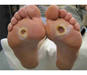
Materials and methods. The study involved patients with diabetes type 2 complicated with diabetic foot syndrome neuroischemic form and the presence of chronic wounds of the lower extremities that do not heal for more than 21 days from the date of occurrence. The study included 31 patients, 7 female and 8 male in the study group and 8 male and female – the control group. Area wounds on the surface of the pressor foot ranged from 3.8 to 21 cm2. The duration of ulcers in patients ranged from 25 days to 3 months. Preparation of ulcers to the plastic closure of the wound defect was carried out as follows. Cleansing the wounds was performed by ultrasonic cavitation. On the wound bandage with sorption-based antimicrobial composition of nanosized silica. After 12 hours, the procedure was repeated cavitation. Thus the wound was purified for 2-3 days. On the third day the wound was applied vacuum-assisted closure (VAC-therapy) with a standard negative pressure of 125 mm Hg. The device was applied for 3 or 6 days depending on the area and depth of the wound. Before the plastic closure of the wound defect was performed bacteriological examination contents wounds. The number of colony-forming units for the success of the operation should be less than 10. Formed flap, which corresponds to the size and configuration of the defect on the foot. Pedicle stands and rotates it in 1800. The donor site is sutured closed or split skin graft. Further, the surface of the flap is superimposed bacteriostatic draining bandage. On the shin and foot removable plaster splint is applied.
Results. In the study group noted engraftment graft in 14 patients (93%). Necrotic flap occurred in one patient due to violations of the immobilization of a limb. Time of full engraftment and the healing of the wound defect in patients of the main group was 14 ± 3 days in the control group healing ulcers – 51 ± 10 days. Long-term observation after 6 months showed complete retention of the supporting limb function and good engraftment, the absence of ulcers on the legs. In the control group after 6 months, 7 patients had recurrent ulcers, one of them has created a need for amputation at projection of Shofar joint.
Conclusions. Improved techniques in the treatment of wound autodermoplastic defects in patients from diabetic foot syndrome, which is the correction of hyperglycemia preparation wounds using a vacuum bandage hardware. Using a split skin graft in conjunction with a vacuum apparatus can effectively cover the bandage chronic and acute wounds, supporting the ability to save limbs and improve the quality of life, reduce the time of hospital stay. Adequate wound closure defects on the foot is a method of preventing high leg amputations in patients with diabetes. Engraftment of skin graft on perforating pedicle accelerated by the use of the method of VAC-therapy.