Інформація призначена тільки для фахівців сфери охорони здоров'я, осіб,
які мають вищу або середню спеціальну медичну освіту.

Підтвердіть, що Ви є фахівцем у сфері охорони здоров'я.


Всесвітній день боротьби із запальними захворюваннями кишечника
день перший
день другий

Коморбідний ендокринологічний пацієнт

Всесвітній день боротьби із запальними захворюваннями кишечника
день перший
день другий

Коморбідний ендокринологічний пацієнт

Международный эндокринологический журнал Том 16, №3, 2020

Вернуться к номеру

The blood level of endothelin-1 in diabetic patients depending on the characteristics of the disease

Авторы: L.K. Sokolova, Yu.B. Belchina, V.V. Pushkarev, S.A. Cherviakova, T.S. Vatseba, O.I. Kovzun, V.M. Pushkarev, M.D. Tronko
SI “V.P. Komisarenko Institute of Endocrinology and Metabolism of the NAMS of Ukraine”, Kyiv, Ukraine

Рубрики: Эндокринология

Разделы: Клинические исследования

Версия для печати


Резюме

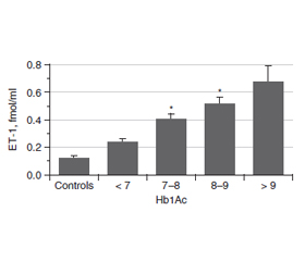
Актуальність. Ендотелін (ЕТ-1) є одним із найбільш значущих регуляторів функціонального стану ендотелію судин. Це найпотужніший вазоконстриктор і маркер ендотеліальної дисфункції. Ендотелій відіграє важливу роль у регуляції тонусу судин. ЕТ-1 чинить як запальну, так і проліферативну дію і призводить до патогенних процесiв у серцево-судинній системі. При цукровому діабеті (ЦД) підвищені концентрації глюкози й глікованого гемоглобіну (HbA1c) впливають на утворення ET-1. Метою роботи було вивчення змісту ЕТ-1 у крові хворих на цукровий діабет із різними показниками індексу маси тіла (ІМТ), тривалістю захворювання й рівнем HbA1c. Матеріали та методи. Концентрацію ЕТ-1 оцінювали методом ІФА у 103 осіб: 17 здорових добровольців і 86 пацієнтів із ЦД. Для визначення ET-1 використовували набір Еndotelin (1-21) EIA kit (1-21) («Biomedica»). HbA1c визначали з використанням набору one HbA1c FS — DiaSys Diagnostic Systems. Результати. Середній рівень ендотеліну в крові хворих на ЦД становив 0,536 ± 0,047 фмоль/мл (конт­роль — 0,118 ± 0,017 фмоль/мл). У всіх пацієнтів із ЦД рівень ET-1 в крові був вищим, ніж у контрольній групі, і збільшувався пропорційно до показника HbA1c. Зі зростанням тривалості ЦД концентрація ET-1 збільшується, досягаючи найвищих значень при тривалості захворювання понад 11 років. Рівень ET-1 у пацієнтів з ожирінням (> 30 кг/м2) значно вищий, ніж у пацієнтів з ІМТ менше від 25 кг/м2 і в діапазоні 25–30 кг/м2. Висновки. Експресія і секреція ET-1 у пацієнтів із цукровим діабетом підвищуються зі збільшенням тривалості захворювання, ІМТ та вмісту HbA1c. Серцево-судинна патологія є основним ускладненням у пацієнтів із ЦД 2-го типу. Ендотеліальна дисфункція належить до ранніх ознак діабетичного судинного ураження. Зміна балансу ЕТ-1 ендотелію є ключовою подією при ініціації атеросклерозу через активацію адгезії лейкоцитів, яка пов’язана з наявністю судинного запалення.

Актуальность. Эндотелин (ЭТ-1) является одним из наиболее значимых регуляторов функционального состояния эндотелия сосудов. Это самый мощный вазоконстриктор и маркер эндотелиальной дисфункции. Эндотелий играет важную роль в регуляции тонуса сосудов. ЭТ-1 оказывает как воспалительное, так и пролиферативное действие и приводит к патогенным процессам в сердечно-сосудистой системе. При диабете повышенные концентрации глюкозы и гликированного гемоглобина (HbA1c) влияют на образование ЭT-1. Целью работы было изучение содержания ЭТ-1 в крови больных сахарным диабетом с различными показателями индекса массы тела (ИМТ), длительности заболевания и уровня HbA1c. Материалы и методы. Концентрация ЭТ-1 оценивалась методом ИФА у 103 человек: 17 здоровых добровольцев и 86 пациентов с сахарным диабетом. Для определения ЭT-1 использовали набор Еndotelin (1-21) EIA kit (1-21) («Biomedica»). Гликированный гемоглобин определяли с использованием набора one HbA1c FS — DiaSys Diagnostic Systems. Результаты. Средний уровень эндотелина в крови больных сахарным диабетом составил 0,536 ± 0,047 фмоль/мл (контроль — 0,118 ± 0,017 фмоль/мл). У всех пациентов с сахарным диабетом уровень ЭT-1 в крови был выше, чем в контрольной группе, и увеличивался пропорционально показателю HbA1c. С увеличением продолжительности сахарного диабета концентрация ЭT-1 возрастает, достигая самых высоких значений при длительности заболевания более 11 лет. Уровень ЭT-1 у пациентов с ожирением (> 30 кг/м2) значительно выше, чем у пациентов с ИМТ менее 25 кг/м2 и в диапазоне 25–30 кг/м2. Выводы. Экспрессия и секреция ЭТ-1 у пациентов с диабетом повышаются с увеличением продолжительности заболевания, ИМТ и содержания HbA1c. Сердечно-сосудистая патология является основным осложнением у пациентов с сахарным диабетом 2-го типа. Эндотелиальная дисфункция относится к ранним признакам диабетического сосудистого поражения. Изменение баланса ЭТ-1 эндотелия является ключевым событием при инициации атеросклероза из-за активации адгезии лейкоцитов, связанной с наличием сосудистого воспаления.

Background. Endothelin (ET-1) is one of the most significant regulators of the functional state of vascular endothelium. It is the most powerful vasoconstrictor and marker for endothelial dysfunction. The endothelium plays an important role in regulating vascular tone. ET-1 has both inflammatory and proliferative effects and contributes to pathogenic processes in the cardiovascular system. In diabetes mellitus (DM), the rise of glucose and glycated hemoglobin (HbA1c) concentration impact the formation of ET-1. The purpose of the work was to study the blood concentration of ET-1 in diabetic patients with various indicators of body mass index (BMI), the duration of the disease and the level of HbA1c. Materials and methods. The concentration of ET-1 was evaluated by ELISA in 103 individuals: 17 healthy volunteers and 86 patients with DM. To determine the ET-1, the endothelin (1-21) EIA kit (Biomedica) was used. Glycated hemoglobin was determined using one HbA1c FS kit — DiaSys Diagnostic Systems. Results. The average blood level of endothelin in patients with DM was 0.536 ± 0.047 fmol/ml (control — 0.118 ± 0.017 fmol/ml). All diabetic patients had blood ET-1 level higher than the control group and it increased in proportion to the amount of HbA1c. With increasing of the DM duration, the ET-1 concentration rises, reaching the highest values with a disease duration > 11 years. ET-1 level in patients with obesity (> 30 kg/m2) is significantly higher than in patients with BMI less than 25 kg/m2 and in the range of 25–30 kg/m2. Conclusions. Thus, the expression and secretion of ET-1 in patients with diabetes mellitus rise up with increasing of the disease duration, BMI and HbA1c content. Cardiovascular morbidity is a major burden in patients with type 2 DM with endothelial dysfunction as an early sign of diabetic vascular disease that is related to the presence of a vascular low-grade inflammation. Alteration in ET-1 balance of the endothelium is the key event in the initiation of arteriosclerosis via activation of leukocyte adhesion, which is linked to the presence of a vascular inflammation.


Ключевые слова

цукровий діабет; ендотелін-1; глікований гемоглобін; ожиріння

сахарный диабет; эндотелин-1; гликированный гемоглобин; ожирение

diabetes mellitus; endothelin-1; glycated hemoglobin; obesity

Introduction

Endothelin (ET-1) is one of the most significant regulators of the vascular endothelium functional state. It is the most powerful vasoconstrictor and marker for endothelial dysfunction (ED). ET-1 is a bicyclic polypeptide consisting of 21 amino acid (Aa) residues formed from the precursor Big-ET-1 under the influence of some enzymes. Its expression mostly occurs in endothelial cells, as well as on the surface of the underlying smooth muscle cells. ET-1 affects receptors of vascular smooth muscle in a paracrine way, causing their contraction and growth, and endothelial cells — in an autocrine-paracrine way, causing the production of vasorelaxants and growth-promoting factors. ET-1 activates some effector systems, including phospholipases C, D, A2, protein kinases, adenylate and guanylate cyclases [1, 2].
The endothelium represents a monolayer of cells covering the lumen of blood vessels. It provides a continuous barrier between blood elements and the artery wall. The endothelium plays an important role in regulating vascular tone by balancing the release of vasodilator factors originating from the endothelium, such as nitric oxide (NO) and prostacyclin, with vasoconstrictor factors, including ET-1, angiotensin II, and reactive oxygen species (ROS) [3].
ET-1 has both inflammatory and proliferative effects and contributes to pathogenic processes in the cardiovascular system. ET-1 counteracts the effects of NO at several levels. It directly inhibits eNOS, decreasing NO release, increases ROS production, activating NADPH oxidase, responsible for the formation of superoxide anion (O2–), which can absorb NO to form peroxynitrite (ONOO–), a powerful free radical that promotes a further decrease in the bioavailability of NO. ET-1 also blocks tetrahydrobiopterin that leads to the uncoupling of eNOS, which begins to produce O2– instead of NO [4].
In diabetes mellitus (DM), the concentration of glucose and glycated hemoglobin (HbA1c) increases, which stimulates the formation of ET-1. The imbalance between the vasodilating and vasoconstrictive actions in the endothelium in this disease is considered to be the most important event in the atherosclerotic process initiation [5, 6].
The purpose of the work was to study the blood content of ET-1 in diabetic patients with various indicators of BMI, the duration of the disease and the level of glycated hemoglobin.

Materials and methods

The amount of ET-1 was evaluated with ELISA in 103 individuals: 17 healthy volunteers and 86 patients with diabetes mellitus. Of these, 16 patients with type 1 DM and 70 — with type 2 DM. The study protocol was approved by the Institute Ethics Committee.
All participants provided written informed consent to the use of their biomaterials for further research. Blood was obtained by standard venipuncture and stored in EDTA vacutainer tubes. Plasma was separated by centrifugation within 10 min after blood sampling. The samples were stored at 80 °C until use. To determine the concentration of ET-1, the endothelin (1-21) EIA kit (Biomedica, Austria) was used. The determination was carried out at an optical density of 450 nm. Glycated hemoglobin was determined using one HbA1c FS kit — DiaSys Diagnostic Systems GmbH (Germany). The measurement was carried out at an optical density of 660 nm.
Statistical calculations and data presentation were performed using Origin 7.0 software. The results of the study are presented as M ± SD. To compare the data groups, Student’s t-test and one-way ANOVA were used. Values of P ≤ 0.05 were considered as significant.
The work is a fragment of scientific research “Epidemiology of cancer in patients with diabetes and the effect of antidiabetic drugs on markers of oncogenesis” (State registration number 0117U005263), which is included in the comprehensive research of Ivano-Frankivsk National Medical University “Оrgans of the respiratory, endocrine, nervous systems in simulated pathological conditions and their correction” (State registration number 0117U001758) without special funding.
The study followed the principles of bioethics: the main provisions of the Council of Europe Convention on Human Rights and Biomedicine (04.04.1997), GCP (1996), the World Medical Association Declaration of Helsinki on the ethical principles of scientific medical research involving human subjects (1964–2000) and the order of the Ministry of Health of Ukraine No 281 from 01.11.2000. All surveyed individuals personally and voluntarily signed an informed consent to participate in the study. The study was approved by the Commission on Biomedical Ethics of the V.P. Komisarenko Institute of Endocrinology and Metabolism of the National Academy of Medical Sciences of Ukraine (Protocol 2 of March 5, 2019).

Results

The average blood level of endothelin in diabetic patients (n = 86) was 0.536 ± 0.047 fmol/ml (control — 0.118 ± 0.017 fmol/ml; n = 17). Differences in the amount of ET-1 in the blood of patients with type 1 and 2 diabetes were insignificant. The dependence of ET-1 concentration on the content of glycated hemoglobin in the blood of patients was most revealing. All patients with diabetes mellitus had blood ET-1 level higher than the control group and it increased in proportion to the amount of HbA1c (Fig. 1). In the blood of patients with HbA1c concentration > 9 %, the amount of ET-1 exceeded the control level by almost 6 times. Differences in the amount of ET-1 between the groups with different HbA1c contents were also observed.
An important indicator of diabetes mellitus is the disease duration. Table 1 shows that even in a relatively short duration of the diabetes mellitus, the amount of ET-1 in the blood of patients increases, reaching the highest values with a disease duration > 11 years.
Another significant parameter, especially for patients with type 2 diabetes, is the body mass index (BMI). ET-1 level in patients with obesity (> 30 kg/m2) is significantly higher than in patients with a BMI of less than 25 kg/m2 and in the range of 25–30 kg/m2 (Table 2).
Thus, the expression and secretion of ET-1 in patients with diabetes rise up with increasing of the disease duration, BMI and glycated hemoglobin content.

Discussion

The molecular mechanisms of the pathological factors influencing ET-1 secretion in diabetes mellitus are not yet fully understood. Synthesis of the biologically active ET-1 peptide (21 Aa residues) is a multistep process. Transcription of the human edn1 gene yields a 2.8-kb mRNA that encodes the 212-Aa preproET-1. A 17-Aa leader sequence targets preproET-1 to the endoplasmic reticulum where it enters the secretory pathway [7]. Prior to exocytosis, furin-like proteases cleave preproET-1 to a protein called big ET-1 (38-Aa). The final cleavage step is mediated by endothelin-converting enzymes that cleave big ET-1 into active ET-1. The regulatory mechanisms obviously exist for each of these post-translational processing steps, however, transcriptional regulation is thought to be the major mechanism controlling ET-1 bioavailability. ET-1 localizes in endothelial cells to both constitutive secretory vesicles and specialized regulatory granules — Weibel-Palade bodies. Hypoxia, thrombin, and shear stress enhance the ET-1 content via exocytosis of Weibel-Palade bodies but are also known to stimulate steady-state edn1 mRNA levels [7]. ET-1 is synthesized and released continuously from endothelial cells, and levels of preproET-1 are modulated predominantly at the level of transcription, with implicating numerous transcription factors including activator protein 1 (AP-1), nuclear factor kappa B (NF-κB), forkhead box protein O1, vascular endothelial zinc finger 1, hypoxia-inducible factor 1, and GATA2. Both physical and chemical stimuli contribute to alterations in levels of preproET-1 mRNA in physiological and pathophysiological conditions [8]. In the vasculature, shear stress is critical in determining the balance between ET-1 and NO production, and alteration in endothelial gene expression appears to involve AMP-activated protein kinase stimulation of the anti-inflammatory transcription factor Krüppel-like factor 2 [9]. Hypoxia also plays an important role in increasing expression of endothelial genes including ET-1 that possess hypoxic responsive elements in their promotors, contributing to disease progression [10]. One of the most important regulators of ET-1 production in endothelial cells is transforming growth factor-β [8].
Obesity generates hypertrophic signals, including ET-1 itself, which stimulate GATA-4 action. The signaling pathway apparently involves activation of RhoA and p38 MAPK and eventually leads to the phosphorylation and activation of GATA-4. Leptin, which is predominantly secreted by adipose cells and whose concentration increases with obesity, also stimulates the expression of ET-1 via JNK, ERK1/2 and the c-Fox/c-Jun heterodimer (AP-1). Fatty acids and lipoproteins enhance the expression of ET-1 through protein kinase C (PKC), which can activate both AP-1 and NF-κB. Finally, low-level chronic inflammation that occurs in obesity and type 2 diabetes, and is associated with the secretion of proinflammatory factors such as tumor necrosis factor-α (TNF-α), interleukin-1β (IL-1β) and interferon-γ (IFN-γ), also activates NF-κB via the PI3K/Akt/IKK signaling pathway [7].
Glycated hemoglobin is known to reflect the blood glucose level, and is a form of hemoglobin which is measured primarily to identify the average plasma glucose concentration over prolonged periods. HbA1c is defined as protein which is irreversibly glycated at one or both N-terminal valines of the beta chains. HbA1c has been the most used and accepted test for monitoring the glycemic control in individuals with diabetes. Once a hemoglobin molecule is glycated, it continues to remain in the red blood cell for the rest of its life-span (~120 days) [11].
Damage action of HbA1c includes an increase of highly reactive free radicals inside blood cells, which alter blood cell membrane properties. This leads to blood cell aggregation and increased blood viscosity, which results in impaired blood flow. Another way HbA1c causes damage is via inflammation, which results in atherosclerotic plaque formation. Free-radical accumulation promotes the increased permeability of endothelium and production of proinflammatory monocyte adhesion proteins, which cause macrophage recruitment on blood vessel surfaces, ultimately leading to formation of plaques in these vessels. Highly glycated Hb-AGEs go through vascular smooth muscle layer and inactivate acetylcholine-induced endothelium-dependent relaxation, possibly through binding to NO, preventing its normal function. NO is a potent vasodilator and it also inhibits formation of plaque-promoting oxidized form of LDL. The degradation of blood cells also releases heme from them that can cause oxidation of endothelial and LDL proteins, which results in plaques [12].
High glucose on its own stimulates the recruitment of NF-κB and p300 to the edn1 promoter and binding of these factors is associated with an increase in histone H3 acetylation [13]. NF-κB is a redox-sensitive transcription factor known to activate edn1 in a variety of cell types [13–15]. The promoter of edn1 contains a functional NF-κB binding site located at position 2090 bp [7]. It is noted that only NF-κB heterodimers (p65/p50) appear to activate edn1 in endothelial cells because coexpression of p65 and p50 subu–nits leads to an increase in ET, whereas expression of p50 alone leads to reduction in edn1 transcription. Recent evidence indicated that the edn1 promoter contained two additional NF-κB binding sites located at 891 and 1214 bp [14]. Stimulation of pulmonary artery smooth muscle cells with TNF-α and IFN-γ led to the binding of NF-κB to all three sites. An increase in regional histone acetylation, a marker of transcriptionally active chromatin, accompanied this NF-κB binding. Several key components of the NF-κB signaling pathway leading to the activation of edn1 have been reported. For example, TNF-α treatment of glioblastoma cells led to association of NF-κB with the edn1 promoter through a pathway that involved PI3K activation. Oleic acid stimulation of edn1 required the activation of calcium-dependent PKC followed by subsequent NF-κB activation [7]. Other cytokines related to obesity are also able to activate NF-κB-dependent edn1 expression. For example, IL-1β treatment resulted in increased NF-κB activation and edn1 expression in renal collecting duct cells [15]. A subcutaneous infusion of IL-1β also resulted in increased renal edn1 expression in mice, and it has been suggested that cytokine-dependent edn1 expression is involved in several in vivo inflammatory processes [17].

Conclusions

Thus, in diabetes mellitus, a high concentration of ET-1 was determined that exceeded normal values. The dependence of the content of this marker on the increase in the disease duration was noted. Even higher concentrations of this peptide in the blood are observed in patients with diabetic micro- and macroangiopathies, which suggests its participation in the development of late vascular complications of diabetes mellitus. High glucose and HbA1c levels as well as obesity increase secretion of ET-1 apparently mainly through stimulation of NF-κB-dependent edn1 expression that causes endothelial dysfunction. A manifestation of ED in diabetes is an impairment of endothelium-dependent vasorelaxation due to a decrease in the production of vasorelaxants and an increased formation of vasoconstrictors, primarily ET-1. Pathogenetically, this mechanism of ED is embodied by activating the polyol pathway of glucose oxidation under conditions of hyperglycemia. Cardiovascular morbidity is a major burden in patients with T2DM with ED as an early sign of diabetic vascular disease that is related to the presence of a vascular low-grade inflammation. Alteration in ET-1 balance of the endothelium is the key event in the initiation of arteriosclerosis, via activation of leukocyte adhesion, which is linked to the presence of a vascular inflammation.
Conflicts of interests. Authors declare the absence of any conflicts of interests and their own financial interest that might be construed to influence the results or interpretation of their manuscript.

Список литературы

  1. Дороднева Е.Ф., Исакова Д.Н., Ефанов А.Ю. Роль эндотелина-1 в развитии кардиоваскулярных заболеваний у пациентов с сахарным диабетом. Медицинская наука и образование Урала. 2013. № 1. С. 172-176.
  2. Дремина Н.Н., Шурыгин М.Г., Шурыгина И.А. Эндотелины в норме и патологии. Международный журнал прикладных и фундаментальных исследований. 2016. № 10–2. С. 210-214.
  3. Tang S.T., Su H., Zhang Q., Tang H.Q., Wang C.J., Zhou Q. et al. Sitagliptin inhibits endothelin-1 expression in the aortic endothelium of rats with streptozotocin-induced diabetes by suppressing the nuclear factor-κB/IκBα system through the activation of AMP-activated protein kinase. Int. J. Mol. Med. 2016. Vol. 37, 6. P. 1558-1566. doi: 10.3892/ijmm.2016.2578.
  4. Maamoun H., Benameur T., Pintus G., Munusamy S., Agouni A. Crosstalk between oxidative stress and endoplasmic reticulum (ER) stress in endothelial dysfunction and aberrant angiogenesis associated with diabetes: a focus on the protective roles of heme oxygenase (HO)-1. Front. Physiol. 2019. Vol. 10. P. 70. doi: 10.3389/fphys.2019.00070.
  5. Тронько Н.Д., Пушкарев В.М., Соколова Л.К., Пушкарев В.В., Ковзун Е.И. Молекулярные механизмы патогенеза сахарного диабета и его осложнений. К.: Издательский дом «Медкнига», 2018. 264 с.
  6. Sokolova L.K., Pushkarev V.M., Pushkarev V.V., Tronko N.D. Diabetes and atherosclerosis. Cellular mechanisms of pathogenesis. Literature review. Endokrynologia. 2017. Vol. 2. P. 127-138.
  7. Stow L.R., Jacobs M.E., Wingo C.S., Cain B.D. Endothelin-1 gene regulation. FASEB J. 2011. Vol. 25. P. 16-28. doi: 10.1096/fj.10-161612.
  8. Davenport A.P., Hyndman K.A., Dhaun N., Southan C., Kohan D.E., Pollock J.S. et al. Endothelin. Pharmacol. Rev. 2016. Vol. 68, № 2. P. 357-418. doi: 10.1124/pr.115.011833.
  9. Young A., Wu W., Sun W., Benjamin Larman H., Wang N., Li Y.-S. et al. Flow activation of AMP-activated protein kinase in vascular endothelium leads to Krüppel-like factor 2 expression. Arterioscler. Thromb. Vasc. Biol. 2009. Vol. 29. P. 1902-1908. doi: 10.1161/ATVBAHA.109.193540.
  10. Spinella F., Caprara V., Cianfrocca R., Rosanò L., Di Castro V., Garrafa E. et al. The interplay between hypoxia, endothelial and melanoma cells regulates vascularization and cell motility through endothelin-1 and vascular endothelial growth factor. Carcinogenesis. 2014. Vol. 35. P. 840-848. doi: 10.1093/carcin/bgu018.
  11. Gupta S., Jain U., Chauhan N. Laboratory diagnosis of HbA1c: a review. J. Nanomed. Res. 2017. Vol. 5, 4. P. 00120. doi: 10.15406/jnmr.2017.05.00120.
  12. Saleh J. Glycated hemoglobin and its spinoffs: Cardiovascular disease markers or risk factors? World J. Cardiol. 2015. Vol. 7, 8. P. 449-453. doi: 10.4330/wjc.v7.i8.449.
  13. Chen S., Feng B., George B., Chakrabarti R., Chen M., Chakrabarti S. Transcriptional co-activator p300 regulates glucose induced gene expression in the endothelial cells. Am. J. Physiol. Endocrinol. Metab. 2010. Vol. 298. P. E127-E137. doi: 10.1152/ajpendo.00432.2009.
  14. Wort S.J., Ito M., Chou P.C., McMaster S.K., Badiger R., Jazrawi E. et al. Synergistic induction of endothelin-1 by tumor necrosis factor alpha and interferon gamma is due to enhanced NF-κB binding and histone acetylation at specific κB sites. J. Biol. Chem. 2009. Vol. 284. P. 24297-24305. doi: 10.1074/jbc.M109.032524.
  15. Boesen E.I., Sasser J.M., Saleh M.A., Potter W.A., Woods M., Warner T.D. et al. Interleukin-1β, but not interleukin-6, enhances renal and systemic endothelin production in vivo. Am. J. Physiol. Renal Physiol. 2008. Vol. 295. P. F446-F453. doi: 10.1152/ajprenal.00095.2008.
  16. El-Mesallamy H., Suwailem S., Hamdy N. Evaluation of C-reactive protein, endothelin-1, adhesion molecule(s), and lipids as inflammatory markers in type 2 diabetes mellitus patients. Mediators Inflamm. 2007. Vol. 2007. P. 73635. doi: 10.1155/2007/73635.
  17. Вацеба Т.С., Соколова Л.К., Пушкарьов В.М. Вплив ожиріння на формування онкологічного ризику у пацієнтів з цукровим діабетом 2-го типу. Міжнародний ендокринологічний журнал. 2020. Т. 16, № 2. С. 90-96. doi: 10.22141/2224-0721.16.2. 2020.201303.

Вернуться к номеру