Журнал «Боль. Суставы. Позвоночник» Том 11, №3, 2021
Вернуться к номеру
Реостеосинтез при псевдоартрозі шийки стегнової кістки
Авторы: Гавриленко Е.О.
КУ «Приморська ЦРЛ», м. Приморськ, Запорізька область, Україна
Рубрики: Ревматология, Травматология и ортопедия
Разделы: Справочник специалиста
Версия для печати
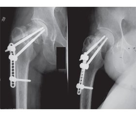
Псевдоартроз шийки стегнової кістки — це тяжке ураження ділянки тазостегнового суглоба, результатом якого в більшості випадків є стійка інвалідизація хворих. Розвиток псевдоартрозу в зазначеному сегменті пояснюється в першу чергу особливостями трофіки проксимального відділу стегнової кістки й поганою якістю кістки. Нормальне функціонування кісткової тканини особливо важливе для швидкого відновлення кістки й остеоінтеграції. Однак, за даними літератури, перелом супроводжується системною втратою кісткової маси на 2–15 % порівняно зі значеннями безпосередньо після перелому або з показниками порівнянних за віком осіб без переломів. Експериментальні дослідження обґрунтовують правомірність використання різних регуляторів метаболізму в кістці для підвищення ефективності загоєння перелому: кальцію, фосфору і протеїнів (колагену, факторів росту, остеокальцину). Наведено опис клінічного випадку відновлення пацієнта після формування псевдоартрозу. У даному випадку була виконана операція реостеосинтезу динамічним фіксатором з кістковою автопластикою шийки правої стегнової кістки. Додатково для покращення кісткової репарації застосовувалися остеотропний комплекс Остеопро® і плазмоліфтинг. Протягом чотирьох місяців після реостеосинтезу була досягнута консолідація перелому, відновлена опорна й рухова функція правої нижньої кінцівки.
Pseudarthrosis of the femoral neck is a severe lesion of the hip joint, the result of which in most cases is a permanent disability of patients. The development of pseudarthrosis in this segment is primarily due to the trophic features of the proximal femur and poor bone quality. Normal functioning of the bone tissue is important for rapid bone regeneration and osseointegration. However, literature data show that the fracture is accompanied by systemic bone loss of 2–15 % compared to the values immediately after the fracture or to the indicators in age-comparable individuals without fractures. Experimental studies substantiate the appropriateness of the use of various regulators of bone metabolism to increase the efficiency of fracture healing: calcium, phosphorus and proteins (collagen, growth factors, osteocalcin). A clinical case of recovery of a patient after the formation of pseudarthrosis is described. In this case, the operation of reosteosynthesis was performed using a dynamic fixator with bone autoplasty of the right femoral neck. Additionally, the osteotropic complex Osteopro® and platelet-rich plasma therapy were used to improve bone repair. Within four months after reosteosynthesis, fracture consolidation was achieved, and the supporting and motor function of the right lower extremity was restored.
псевдоартроз; реостеосинтез; динамічний фіксатор; перелом шийки стегнової кістки; Остеопро®
pseudarthrosis; reosteosynthesis; dynamic fixator; femoral neck fracture; Osteopro®
- Kavalerskiy G.M., Murylyov V.Yu., Rubin G.G., Rukin Ya.A., Elizarov P.M., Muzychenko A.V. Hip arthroplasty in patients with femoral neck pseudoarthrosis. Herald of Traumatology and Orthopedics. N.I. Priorov. 2016. 1. 21-6.
- Егиазарян К.А., Сиротин И.В., Бут-Гусаим А.Б., Горбачев М.А. Псевдоартроз шейки бедра: особенности возникновения и тактики лечения. Российский медицинский журнал. 2018. 24(4). 195-198. DOI: http://dx.doi.org/10.18821/0869-2106-2018-24-4-195-198
- Hitt K. Femoral neck nonunion: osteotomy or arthroplasty. Tech. Orthop. 2002. 17 434-442. doi: 10.1097/00013611-200212000-00007.
- Fox K.M., Magaziner J., Hawkes W.G., Yu-Yahiro J., Hebel J.R., Zimmerman S.I. et al. Loss of bone density and lean body mass after hip fracture. Osteoporos. Int. 2000. 11(1). 31-5. DOI: 10.1007/ s001980050003.
- Karlsson M.K., Josefsson P.O., Nordkvist A., Akesson K., Seeman E., Obrant K.J. Bone loss following tibial osteotomy: a model for evaluating posttraumatic osteopenia. Osteoporos. Int. 2000. 11. 261-4.
- Ahmed L.A., Center J.R., Bjørnerem A., Bluic D., Joakimsen R.M., Jørgensen L. et al. Progressively increasing fracture risk with advancing age after initial incident fragility fracture: the Tromsø study. J. Bone Miner. Res. 2013. 28(10). 2214-21. DOI: 10.1002/jbmr.1952.
- Lyles K.W., Schenck A.P., Colón-Emeric C.S. Hip and other osteoporotic fractures increase the risk of subsequent fractures in nursing home residents. Osteoporos. Int. 2008. 19(8). 1225-33. DOI: 10.1007/s00198-008-0569-3.
- Лунева С.Н., Матвеева Е.Л., Гасанова А.Г., Бойчук С.П., Сазонова Н.В. Роль кальция и витамина D3 в восстановлении целостности костей после переломов. Доктор.Ру. 2019. № 2(157). С. 55-60. DOI: 10.31550/1727-2378-2019-157-2-55-60.
- Einhorn T.A., Gerstenfeld L.C. Fracture healing: mechanisms and interventions. Nat. Rev. Rheumatol. 2015. 11(1). 45-54. DOI: 10.1038/nrrheum.2014.164.
- Nikolaou V.S., Efstathopoulos N., Kontakis G., Kanakaris N.K., Giannoudis P.V. The influence of osteoporosis in femoral fracture healing time. Injury. 2009. 40(6). 663-8. DOI: 10.1016/j. injury.2008.10.035.
- Castelo-Branco C. Management of osteoporosis. An overview. Drugs Aging. 1998. Vol. 12. P. 25-32.


/44.jpg)
/45.jpg)