
Журнал «Здоровье ребенка» Том 17, №6, 2022
Вернуться к номеру
Оцінка забезпечення селеном у підлітків, які проживають у сільській місцевості
Авторы: Гончарова О.А. (1), Паньків В.І. (2), Паньків І.В. (3)
(1) — Харківський національний медичний університет, м. Харків, Україна
(2) — Український науково-практичний центр ендокринної хірургії, трансплантації ендокринних органів і тканин МОЗ України, м. Київ, Україна
(3) — Буковинський державний медичний університет, м. Чернівці, Україна
Рубрики: Педиатрия/Неонатология
Разделы: Клинические исследования
Версия для печати
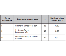
Вступ. Установлено, що дефіцит селену є одним із чинників ризику розвитку тиреоїдної патології, у тому числі автоімунної. Доказова база цього твердження містить дані стосовно поєднання низького рівня селену в організмі зі збільшенням об’єму щитоподібної залози (ЩЗ), гетерогенністю та наявністю гіпоехогенності, які є ультрасонографічними ознаками тиреоїдної лімфоїдної інфільтрації. З огляду на те, що мешканці сільської місцевості вживають переважно овочі та фрукти місцевого походження та воду з місцевих джерел, рівні селену в них значною мірою відображають забезпеченість цим мікроелементом на конкретних територіях. Мета. Визначити ступінь забезпеченості селеном дівчаток-підлітків сільської місцевості Запорізької та Харківської областей України. Матеріали та методи. Досліджено рівень селену у волоссі дівчаток-підлітків віком 13–15 років. Вимірювання проведено за допомогою атомно-абсорбційного спектрометра ICE 3500 (Thermo Fisher Scientific, США). За нормальні показники приймали вміст селену понад 0,8 мкг/г. Результати. Встановлено, що вміст селену у волоссі обстежених дівчаток був нижчий від референтних значень у 15 із 16 мешканок Запорізької області (93,6 %), у 16 із 18 — Балаклійського району (88,9 %) та в усіх обстежених дівчаток із Чугуївського району Харківської області (100,0 %). При цьому рівні медіани і мінімальних значень забезпеченості селеном в усіх групах обстежених були меншими від нижчого порогу референтних норм щодо забезпеченості селеном. У 12 з 16 обстежених І групи (75,0 %), 9 з 12 обстежених ІІ групи (75,0 %) та 14 з 18 обстежених ІІІ групи (77,77 %) розміри ЩЗ перевищували вікову норму. Висновки. Результати проведеного дослідження свідчать, що в дівчаток-підлітків, які мешкають на сільських територіях Запорізької і Харківської областей, спостерігається висока частота дефіциту селену, на тлі якого у більшості з обстеженого контингенту відзначається збільшення об’єму ЩЗ. Такі дані потребують забезпечення регулярного контролю за станом здоров’я підлітків вказаних місцевостей із обов’язковою участю ендокринологів. Крім того, є потреба в розробці особливих організаційних заходів, які б забезпечили своєчасне виявлення дітей із дефіцитом селену з раціональним графіком їх подальшого обстеження.
Background. It has been found that selenium deficiency is one of the risk factors for the development of thyroid pathology, in particular autoimmune one. The evidence base for this statement includes the association of low selenium levels with enlarged thyroid gland, heterogeneity, and the presence of hypoechogenicity, which are ultrasonographic signs of thyroid lymphoid infiltration. Given that the inhabitants of rural areas consume mainly vegetables and fruits of local origin and water from local sources, the levels of selenium in them largely reflect the supply of this trace element in specific territories. Aim: to determine the degree of selenium availability of adolescent girls in rural areas of Zaporizhzhia and Kharkiv regions of Ukraine. Materials and methods. The level of selenium was studied in the hair of adolescent girls aged 13–15 years. Measurements were made using an atomic absorption spectrometer ICE 3500 (Thermo Fisher Scientific, USA). Selenium content of more than 0.8 μg/g was considered normal. Results. It was found that selenium level in the hair was lower than the reference values in 15 of 16 residents of Zaporizhzhia region (93.6 %), in 16 of 18 residents of the Balakliia district (88.9 %) and in all the examined girls from Chuhuiiv district of Kharkiv region (100.0 %). At the same time, the median and minimum levels of selenium supply in all groups of patients were lower than the lower threshold of reference norms. In 12 of 16 examinees in group I (75.0 %), 9 of 12 in group II (75.0 %) and 14 of 18 in group III (77.77 %), thyroid sizes exceeded the age norm. Conclusions. The results of the research show that adolescent girls living in rural areas of Zaporizhzhia and Kharkiv regions have a high frequency of selenium deficiency against the background of which the majority of the examined patients has increased thyroid volume. Such data require regular monitoring of the health of adolescents in the specified areas with the mandatory participation of endocrinologists. In addition, there is a need to develop special organizational measures that would ensure timely detection of children with selenium deficiency with a rational schedule for their further examination.
селен, щитоподібна залоза, забезпечення, діти, сільське населення
selenium; thyroid gland; supply; children; rural population
Вступ
Матеріали та методи
Результати
/33.jpg)
/34.jpg)
Обговорення
Висновки
- Winther K.H., Rayman M.P., Bonnema S.J., Hegedüs L. Selenium in thyroid disorders — essential knowledge for clinicians. Nature reviews. Endocrinology. 2020. 16(3). 165-176. DOI: 10.1038/s41574-019-0311-6. PMID: 32001830.
- Kravchenko V.I., Grossman A.B., Rakov O.V., Kovzun O.I., Pankiv V.I., Simurov O.V. Selenium supply and thyroid condition in Graves’ disease in the region of iodine deficiency. Problems of Endocrine Pathology. 2021. 1. 26-33. https://doi.org/10.21856/j-PEP.2021.1.04.
- Kobayashi R., Hasegawa M., Kawaguchi C., Ishikawa N., Tomiwa K., Shima M., Nogami K. Thyroid function in patients with selenium deficiency exhibits high free T4 to T3 ratio. Clin. Pediatr. Endocrinol. 2021. 30(1). 19-26. doi: 10.1297/cpe.30.19.
- Hu Y., Feng W., Chen H., Shi H., Jiang L., Zheng X., Liu X., et al. Effect of selenium on thyroid autoimmunity and regulatory T cells in patients with Hashimoto’s thyroiditis: A prospective randomized-controlled trial. Clin. Transl. Sci. 2021. 14(4). 1390-1402. doi: 10.1111/cts.12993.
- Goncharova O.A., Karachentsev Yu.I. Autoimmune thyroid disease. K.: Publisher Zaslavsky A.Yu., 2017. 212 p. (in Russian)
- Shidlovsky V.O., Pankiv V.I. Autoimmune thyroiditis in the new reality. Ternopil: TNMU, 2021. 278 p. (in Ukrainian)
- Pankiv I. Prevalence of autoimmune thyroiditis among wo–men with vitamin D deficiency. International Journal of Endocrinology (Ukraine). 2017. 13(5). 336-339. https://doi.org/10.22141/2224-0721.13.5.2017.110023 (in Ukrainian)
- Hubalewska-Dydejczyk A., Duntas L., Gilis-Januszewska A. Pregnancy, thyroid, and the potential use of selenium. Hormones (Athens). 2020. 19(1). 47-53. doi: 10.1007/s42000-019-00144-2.
- Tortelly Costa V.D., Melo D.F., Matsunaga A.M. The Relevance of Selenium to Alopecias. Int. J. Trichology. 2018. 10(2). 92-93. doi: 10.4103/ijt.ijt_37_17. PMID: 29769785; PMCID: PMC5939011.
- Ventura M., Melo M., Carrilho F. Selenium and Thyroid Disease: From Pathophysiology to Treatment. Int. J. Endocrinol. 2017. 2017. 1297658. doi: 10.1155/2017/1297658. PMID: 28255299; PMCID: PMC5307254.
- García-Ascaso M.T., Ares Segura S., Ros Pérez P., Piñeiro Pérez R., Alfageme Zubillaga M. Thyroid Volume Assessment in 3-14 Year-Old Spanish Children from an Iodine-Replete Area. Eur. Thyroid J. 2019. 8(4). 196-201. doi: 10.1159/000499103. PMID: 31602362; PMCID: PMC6738148.
- Köhrle J. Selenium and the thyroid. Curr. Opin. Endocrinol. Diabetes Obes. 2015. 22(5). 392-401. doi: 10.1097/MED.0000000000000190. PMID: 26313901.
- Pashkovska N. Selenium and autoimmune thyroid disorders. International Journal of Endocrinology (Ukraine). 2017. 13(1). 33-38. https://doi.org/10.22141/2224-0721.13.1.2017.96753.
- Szeliga A., Czyżyk A., Niedzielski P., Mleczek M., Maciejewski A., Dorszewska J., Łącka K. Assessment of serum selenium concentration in patients with autoimmune thyroiditis in Poznan district. Pol. Merkur. Lekarski. 2018. 45(268). 150-153. PMID: 30371648.
- Lönnerdal B., Vargas-Fernández E., Whitacre M. Selenium fortification of infant formulas: does selenium form matter? Food Funct. 2017. 8(11). 3856-3868. doi: 10.1039/c7fo00746a. PMID: 28991311.
- Stoffaneller R., Morse N.L. A review of dietary selenium intake and selenium status in Europe and the Middle East. Nutrients. 2015. 7(3). 1494-537. doi: 10.3390/nu7031494. PMID: 25734564; PMCID: PMC4377864.
- Filonenko M., Zhuravlyova L., Sokolnikova N. Correlation of cardiac biomarkers with the levels of selenium and antioxidant enzymes in patients with acute myocardial infarction and a history of hypertension. Wiad. Lek. 2022. 75(2). 362-365. PMID: 35307659.
- Stabnikova O., Antoniuk M., Stabnikov V., Arsen’eva L. Ukrainian Dietary Bread with Selenium-Enriched Soya Malt. Plant Foods Hum. Nutr. 2019. 74(2). 157-163. doi: 10.1007/s11130-019-00731-z. PMID: 31020517.
- Tarashchenko Yu.M., Kovalenko A.E., Kravchenko V.I., Kovzun O.I., Simurov O.V. Iodine and selenium deficiency in the pathogenesis of thyroid goiter transformation and thyroid autoimmune disorders (literature review and results of own research). Endokrynologia. 2020. 25 (4). 297-304. DOI: 10.31793/1680-1466.2020.25-4.297 (in Ukrainian).