Журнал «Боль. Суставы. Позвоночник» Том 12, №2, 2022
Вернуться к номеру
Вплив ходьби на формування кульшового суглоба в пацієнтів з дитячим церебральним паралічем
Авторы: Яцуляк М., Марциняк С., Філіпчук В.
ДУ «Інститут травматології та ортопедії НАМН України», м. Київ, Україна
Рубрики: Ревматология, Травматология и ортопедия
Разделы: Клинические исследования
Версия для печати
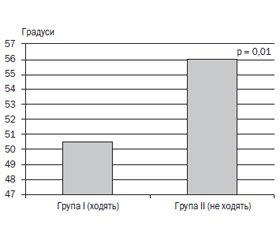
Актуальність. Вплив ходьби на формування кульшового суглоба в пацієнтів з дитячим церебральним паралічем (ДЦП) є актуальним об’єктом наукових досліджень. Мета дослідження: встановити залежність між ходьбою і клініко-рентгенограмометричними параметрами (КРМП) кульшового суглоба в дітей із ДЦП. Матеріали та методи. Загальна кількість обстежених становила 39 осіб із ДЦП і патологією кульшових суглобів (70 суглобів), які лікувались у ДУ «Інститут травматології та ортопедії НАМН України» за період з 2018 по 2022 рік. Для аналізу хворі були поділені на дві групи залежно від можливості ходьби. Нами проведено клініко-рентгенограмометричне обстеження кульшових суглобів власним способом і їх стандартне передньо-заднє рентгенологічне дослідження. Усім пацієнтам виконували клінічну оцінку торсії стегнової кістки за методикою Ruwe. Для оцінки зв’язків між отриманими показниками виконано кореляційний аналіз Спірмена. Результати. Виявлено позитивний вплив ходьби на формування кульшового суглоба в осіб із ДЦП. Середні значення КРМП кульшових суглобів у пацієнтів із ДЦП, які не ходять, були вірогідно вищими порівняно з показниками осіб, які ходять. Кореляційний аналіз дозволив виявити зв’язки між ходьбою і досліджуваними параметрами кульшового суглоба (шийково-діафізарний кут, торсія стегнової кістки, ацетабулярний кут, кут Шарпа, індекс Реймерса, кут Віберга) в обох укладках, а також між ходьбою та іншими факторами (вік, рівень ураження, показник за шкалою GMFCS (ІІ–ІV рівень), уроджена дисплазія кульшових суглобів в анамнезі). При цьому не знайдено залежності між ходьбою і міотомією аддукторів в анамнезі. Висновки. Встановлено вірогідний вплив функції ходьби на наступні параметри кульшового суглоба: істинний шийково-діафізарний кут (p = 0,00001) в укладці власним способом, торсія стегнової кістки (p = 0,01), ацетабулярний кут (стандартна укладка) (p = 0,00001), кут Шарпа (стандартна укладка) (p = 0,018), індекс Реймерса (стандартна укладка) (p = 0,00007), кут Віберга (стандартна укладка) (p = 0,001) — і відсутність статистичної значущості впливу фактора міотомії аддукторів в анамнезі (p = 0,11) на функцію ходьби.
Introduction. The influence of the gait on the hip joint formation in patients with infantile cerebral palsy (CP) is an actual object of scientific research. The purpose of the research was to study the correlations between walking and clinical and roentgenometric parameters of the hip joint in the patients with CP. Materials and methods. There were examined 39 patients with CP and pathology of the hip joints (70 joints), who had been treated at the National Research Institute of Traumatology and Orthopedics for the period from 2018 to 2022. The patients had been divided into 2 groups depending on the ability to walk. We had performed a clinical and roentgenogramometric examination of the hip joints in positioning according to our own method and the standard anterior-posterior radiological position. All patients underwent a clinical assessment of femoral torsion using the Ruwe method. To assess the relationships between the studied indices Spearman's correlation analysis was performed. Results. The positive influence of gait on the hip joint formation had been revealed. The average values of the hip clinical and roentgenometric parameters in patients with CP who do not walk were significantly higher compared to those of subjects who walk. Correlation analysis revealed the relationship between walking and the studied parameters of the hip joint (cervical-diaphyseal angle, femoral torsion, acetabular angle, Sharp angle, Reimers index, the center edge angle of Wiberg) in both settings, as well as walking and other factors (age, level lesion, GMFCS scale (II-IV level), congenital dysplasia of the hip joints in the history). At the same time, there was no found any dependence between gait and adductor myotomy in the history. Conclusions. A significant influence of the gait on the following parameters of the hip joint was established: the true cervical-diaphyseal angle (p = 0.00001) in positioning according to our own method, femoral torsion (p = 0.01), acetabular angle (standard setting) (p = 0.00001), Sharpe angle (standard setting) (p = 0.018), Reimers index (standard setting) (p = 0.00007), center edge angle of Wiberg (standard setting) (p = 0.001) and lack of statistical significance of the influence of the adductor myotomy factor in history (p = 0.11) on the walking function.
дитячий церебральний параліч; кульшовий суглоб; рентгенограмометричні показники; ходьба; індекс Реймерса
infantile cerebral palsy; hip joint; roentgenogrammetric indicators; gait; Reimers index
Вступ
Матеріали та методи
Результати
Обговорення
Висновки
- Bosmans L., Jansen K., Wesseling M., Molenaers G., Scheys L., Jonkers I. The role of altered proximal femoral geometry in impaired pelvis stability and hip control during CP gait: A simulation study. Gait Posture. 2016. 44. 61-7. doi: 10.1016/j.gaitpost.2015.11.010. Epub 2015 Nov 30. PMID: 27004634.
- Yatsuliak M., Nemesh M., Filipchuk V. Factors influencing the formation of the proximal femur in patients with cerebral palsy. Wiad. Lek. 2022. 75(6). 1642-1647. doi: 10.36740/wlek202207105. PMID: 35962673.
- Upadhyay S.S., Burwell R.G., Moulton A., Small P.G., Wallace W.A. Femoral anteversion in healthy children. Application of a new method using ultrasound. J. Anat. 1990. 169. 49-61. PMID: 2200768; PMCID: PMC1256956.
- Palisano R., Rosenbaum P., Walter S., Russell D., Wood E., Galuppi B. Development and reliability of a system to classify gross motor function in children with cerebral palsy. Dev. Med. Child Neurol. 1997. 39(4). 214-223. doi: 10.1111/j.1469-8749.1997.tb07414.x. PMID: 9183258.
- Куценок Я.Б., Рулла Е.А., Мельник В.В. Вроджена дисплазія кульшового суглоба. Вроджений підвивих і вивих стегна. Київ: Здоров’я, 1992. 184 p.
- Reimers J. The stability of the hip in children. A radiological study of the results of muscle surgery in cerebral palsy. Acta Orthop. Scand. Suppl. 1980. 184. 1-100. doi: 10.3109/ort.1980.51.suppl-184.01. PMID: 6930145.
- Ruwe P.A., Gage J.R., Ozonoff M.B., DeLuca P.A. Clinical determination of femoral anteversion. A comparison with established techniques. J. Bone Joint Surg. Am. 1992. 74(6). 820-30. PMID: 1634572.
- Гошко В.Ю., Науменко Н.О., Чеверда А.І., Яцуляк М.Б., Немеш М.М., винахідники; ДУ «Інститут травматології та ортопедії НАМН України», патентовласник. Спосіб визначення клініко-рентгенограмометричних показників кульшового суглоба у пацієнтів з патологією кульшового суглоба. Патент України № 137567. 2019 жов. 25.
- Гошко В.Ю., Науменко Н.О., Яцуляк М.Б., Чеверда А.І., Немеш М.М., Марциняк С.М. Обґрунтування способу визначення клініко-рентгенограмометричних показників кульшового суглоба в пацієнтів із ДЦП. Травма. 2021. 22(1). 61-65 doi: 10.22141/1608-1706.1.22.2021.226411.
- Yatsulіak M., Nemesh M., Martsyniak S., Kabatsii M., Filipchuk V. Original positioning method to determine the clinical and radiographic parameters of the hip joint in patients with cerebral palsy. MOJ Orthopedics & Rheumatology. 2021 Aug 13(4). 90-93. doi: 10.15406/mojor.2021.13.00555. Режим доступу: https://medcraveonline.com/MOJOR/MOJOR-13-00555.pdf.
- Гошко В.Ю., Науменко Н.О., Яцуляк М.Б., винахідники; ДУ «Інститут травматології та ортопедії НАМН України», Гошко В.Ю., Яцуляк М.Б., патентовласники. Ортопедична приставка для укладання пацієнта при рентгенографії кульшових суглобів. Патент України на винахід № 122629. 2020 груд. 10.
- Kim H.Y., Cha Y.H., Byun J.Y., Chun Y.S., Choy W.S. Changes in gait parameters after femoral derotational osteotomy in cerebral palsy patients with medial femoral torsion. J. Pediatr. Orthop. B. 2018. 27(3). 194-199. doi: 10.1097/BPB.0000000000000467. PMID: 28537994; PMCID: PMC5895112.
- Galarraga C.O.A., Vigneron V., Dorizzi B., Khouri N., Desailly E. Predicting postoperative gait in cerebral palsy. Gait Posture. 2017. 52. 45-51. doi: 10.1016/j.gaitpost.2016.11.012. Epub 2016 Nov 9. PMID: 27871017.
- Desailly E., Badina A., Khouri N. Kinematics after unilateral femoral derotation osteotomy in children with diplegic cerebral palsy. Orthop. Traumatol. Surg. Res. 2020. 106(7). 1325-1331. doi: 10.1016/j.otsr.2019.11.032. Epub 2020 Apr 29. PMID: 32360555.
- Park B.S., Chung C.Y., Park M.S., Lee K.M., Cho S.H., Sung K.H. Effects of soft tissue surgery on transverse kinematics in patients with cerebral palsy. BMC Musculoskelet. Disord. 2019. 20(1). 566. doi: 10.1186/s12891-019-2955-8. PMID: 31775715; PMCID: PMC6882030.
- de Morais Filho M.C., Blumetti F.C., Kawamura C.M., Ferreira C.L. Jr, Lopes J.A.F., Fujino M.H., Neves D.L. The effect of the Majestro-Frost procedure on internal hip rotation during gait in patients with cerebral palsy. Gait Posture. 2018. 66. 32-37. doi: 10.1016/j.gaitpost.2018.08.014. Epub 2018 Aug 18. PMID: 30142452.
- Schwarze M., Block J., Kunz T., Alimusaj M., Heitzmann D.W.W., Putz C., Dreher T., Wolf S.I. The added value of orthotic management in the context of multi-level surgery in children with cerebral palsy. Gait Posture. 2019. 68. 525-530. doi: 10.1016/j.gaitpost.2019.01.006. Epub 2019 Jan 6. PMID: 30623847.
- Rose J., Cahill-Rowley K., Butler E.E. Artificial Walking Technologies to Improve Gait in Cerebral Palsy: Multichannel Neuromuscular Stimulation. Artif. Organs. 2017. 41(11). E233-E239. doi: 10.1111/aor.13058. PMID: 29148138.
- Greve K.R., Joseph C.F., Berry B.E., Schadl K., Rose J. Neuromuscular electrical stimulation to augment lower limb exercise and mobility in individuals with spastic cerebral palsy: A scoping review. Front. Physiol. 2022. 13. 951899. doi: 10.3389/fphys.2022.951899. PMID: 36111153; PMCID: PMC9468780.
- Matsuda M., Iwasaki N., Mataki Y., Mutsuzaki H., Yoshikawa K., Takahashi K. et al. Robot-assisted training using Hybrid Assistive Limb® for cerebral palsy. Brain Dev. 2018. 40(8). 642-648. doi: 10.1016/j.braindev.2018.04.004. Epub 2018 Jul 14. PMID: 29773349.


/24.jpg)
/25.jpg)
/26.jpg)
/26_2.jpg)
/27.jpg)
/27_2.jpg)