
Журнал «Здоровье ребенка» Том 17, №8, 2022
Вернуться к номеру
Стан кальцієвого гомеостазу і окремі аспекти його порушень при ювенільному ідіопатичному артриті
Авторы: Антипкін Ю.Г. (1), Марушко Ю.В. (2), Омельченко Л.І. (1), Муквіч О.М. (1), Людвік Т.А. (2), Бондаренко Н.Ю. (1), Бовкун О.А. (2), Ісмакаєва Д.Л. (1)
(1) — ДУ «Інститут педіатрії, акушерства і гінекології ім. акад. О.М. Лук’янової НАМН України», м. Київ, Україна
(2) — Національний медичний університет ім. О.О. Богомольця, м. Київ, Україна
Рубрики: Педиатрия/Неонатология
Разделы: Клинические исследования
Версия для печати
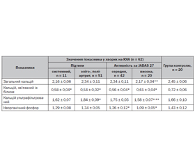
Актуальність. У патогенезі ювенільного ідіопатичного артриту (ЮІА) головну роль відіграють імунопатологічні зміни в організмі з втратою толерантності до елементів власних тканин, при цьому важливе значення мають порушення в обміні кальцію (Са) та метаболізмі кісткової тканини, які виникають внаслідок автоімунного запалення, застосованої фармакотерапії та дії низки інших чинників, що негативно впливають на кальцієвий гомеостаз в організмі. Мета дослідження: дослідити особливості кальцієвого гомеостазу й окремі аспекти його порушень з оцінкою структурно-функціонального стану кісткової тканини при ЮІА з урахуванням клінічних підтипів і активності захворювання. Матеріали та методи дослідження. Обстежено 62 дитини з ЮІА, з них 11 із системним і 51 пацієнт з оліго- та поліартритом віком від 3,5 до 16 років. Визначали концентрації у сироватці крові загального кальцію (за допомогою тест-набору Lachema, Чехія), зв’язаної з білком та ультрафільтрованої фракцій, вміст неорганічного фосфору (загальноприйнятим спектрофотометричним методом за допомогою аналізатора Cobas 6000 з використанням тест-системи Roche Diagnostics, Швейцарія), активність загальної лужної фосфатази (ЛФ) і активність її ізоферментів (кісткової та кишкової) з використанням тест-системи Lachema (Чехія). Проводили ультразвукову остеометрію п’яткової (трабекулярної) кістки за допомогою апарата Achilles фірми Lunar (США). Результати. Встановлено вірогідне зменшення середньої концентрації загального кальцію, кальцію, зв’язаного з білком, при системному ЮІА та при високій активності захворювання. Концентрація ультрафільтрованої фракції кальцію знижувалася лише при високій активності хвороби. Середня концентрація неорганічного фосфору в дітей з оліго- та поліартритами перебувала в межах норми, а при системному ЮІА — знижувалася. Вірогідне зниження вмісту неорганічного фосфору в сироватці крові відмічено у пацієнтів з високою активністю системного ЮІА. При високій активності хвороби у дітей із системним ЮІА виявляється вірогідне зниження показників активності загальної ЛФ та її кісткового ізоферменту. У хворих на ЮІА (оліго-, поліартрит) з повільно прогресуючим перебігом ревматичного процесу протягом першого року захворювання вірогідно змінювався лише показник широкосмугового ослаблення ультразвуку, швидкість поширення ультразвуку та індекс міцності кісткової тканини не змінювалися. У пацієнтів з більшою тривалістю хвороби суттєво зменшувалися всі денситометричні показники. У групі хворих із системним ЮІА та швидко прогресуючим перебігом, високою активністю захворювання, що потребувало глюкокортикоїдної терапії, значна втрата кісткової маси відзначалася вже наприкінці 1-го року захворювання. Висновки. При ЮІА мають місце зміни в показниках концентрації загального кальцію та його білковозв’язаної та ультрафільтрованої фракції у сироватці крові, які свідчать про напруженість кальцій-фосфорного обміну та можливий дефіцит кальцію в організмі вже на ранніх стадіях патологічного процесу. Зниження активності ферменту лужної фосфатази та її кісткового ізоферменту асоціюється з порушеннями структурно-функціонального стану кісткової системи у хворих на ЮІА, що прогресують із тривалістю хвороби. Хворі з ЮІА потребують своєчасної діагностики та моніторингу порушень кальцій-фосфорного обміну з оцінкою структурно-функціонального стану кісткової системи для цілеспрямованої корекції комплексної терапії за рахунок застосування препаратів для підвищення процесів регенерації кісткової тканини та зменшення прогресування остеопенії й остеопорозу і збереження здоров’я зростаючого організму.
Background. In the pathogenesis of juvenile idiopathic arthritis (JIA), the main role is played by immunopathological changes in the body with a loss of tolerance to the elements of own tissues; herewith, disorders of calcium and bone metabolism are very important. Such changes occur as a result of autoimmune inflammation, pharmacotherapy, and the influence of a number of other factors that negatively affect calcium homeostasis in the body. Purpose: to study the features of calcium homeostasis and certain aspects of its disorders with an assessment of the structural and functional state of bone tissue, taking into account clinical subtypes and disease activity. Material and methods. Sixty-two children with JIA aged 3.5 to 16 years were examined, of them 11 had systemic and 51 had oligo- and polyarthritis. There were determined serum concentrations of a total calcium using the Lachema test kit (Czech Republic), protein-bound and ultrafiltered fractions, content of inorganic phosphorus (with the generally accepted spectrophotometric method using the Cobas 6000 analyzer and test systems by Roche Diagnostics, Switzerland), the activity of total alkaline phosphatase and its isoenzymes (bone and intestinal) using the Lachema test system (Czech Republic). Ultrasonic osteometry of the calcaneal (trabecular) bone was performed on the Achilles device (Lunar, USA). Results. A significant decrease was found in the average concentration of total calcium, protein-bound calcium in systemic JIA and in high disease activity. The concentration of the ultrafiltered calcium fraction decreased only with high disease activity. The average concentration of inorganic phosphorus in children with oligo- and polyarthritis was within the normal range, while in systemic JIA it decreased. A significant decrease in the serum content of inorganic phosphorus, as well as in the activity of total alkaline phosphatase and its bone isoenzyme was detected in patients with high activity of systemic JIA. In patients with JIA (oligo-, polyarthritis) characterized by a slowly progressive rheumatic process, only the indicator of broadband ultrasound attenuation significantly changed during the first year of the disease, while the speed of ultrasound propagation and the index of bone tissue strength were not changed. In patients with a longer duration of the disease, all densitometric indicators decreased significantly. In the group of patients with systemic JIA and a rapidly progressive course, high activity of the disease that required a glucocorticoid therapy, a significant loss of bone mass was noted by the end of the first year of the disease. Conclusions. In JIA, there are changes in the concentration of total calcium and its protein-bound and ultrafiltered fractions in the blood serum, which indicate the tension of calcium-phosphorus metabolism and possible calcium deficiency in the body already at the early stages of the pathological process. A decrease in the activity of the alkaline phosphatase and its bone isoenzyme is associated with a violation of the structural and functional changes in the bone system of patients with JIA, which progresses with the duration of the disease. Patients with JIA require timely diagnosis and monitoring of calcium-phosphorus metabolism disorders with an assessment of the structural and functional state of the bone system for purposeful correction of comprehensive therapy due to the use of drugs in order to increase bone tissue regeneration, reduce the progression of osteopenia and osteoporosis, and preserve the health of the growing organism.
ювенільний ідіопатичний артрит; кальцій; лужна фосфатаза; вітамін D; діти
juvenile idiopathic arthritis; calcium; alkaline phosphatase; vitamin D; children
Для ознакомления с полным содержанием статьи необходимо оформить подписку на журнал.
- Sheila T., Angeles-Han., Sarah Ringold, et al. American College of Rheumatology. Arthritis Foundation Guideline for the Screening, Monitoring, and Treatment of Juvenile Idiopathic Arthritis-Associated Uveitis. Arthritis Care & Research. Vol. 71. No. 6. June 2019. Р. 703-716.
- Шевченко Н.С., Богмат Л.Ф., Хаджинова Ю.В. Стан кісткової тканини в дітей з ювенільним ідіопатичним артритом. Сучасна педіатрія. Україна. 2021. № 1(113). С. 45-52. doi: 10.15574/SP. 2021.113.45.
- James L., Lewis III, MD Brookwood Baptist Health and Saint Vincent’s Ascension Health, Birmingham, oct. 2021.
- Sufia Khatun Sumi, Shahana Rahman, Mohammad Immul Islam, Mohammad Mahbubul Islam, Manik Kumar Taluder. Assessment of vitamin D, Calcium, inorganic phosphate, alkaline phosphatase and parathormone in juveile idiopathic arthritis patients Bangladesh S. Child Health. 2019. Vol. 43(3). 145-151.
- Поворознюк В.В., Віленський А.Б., Григор’єва Н.В. Остеопенічний синдром у дітей та підлітків: фактори ризику, діагностика, профілактика. Методичний посібник. Київ, 2001. 28 с.
- Марушко Т.В., Голубовська Ю.Є. Чи можливо передбачити остеопенію у хворих на ювенільний ідіопатичний артрит? Здоров’я дитини. 2019. Т. 14. № 7.
- Calcium requirements in adolescents. Steven A. Abrams, MD. All topics are updated as new evidence becomes available and our peer review process is complete. Literature review current through: Aug. 2022. This topic last updated: Feb II, 2021.
- Lisakovska O., Shymanskyi I., Labudzynskyi D., Mazanova A., Veliky M. Vitamin D Auto-/Paracrine System Is Involved in Modulation of Glucocorticoid-Induced Changes in Angiogenesis/Bone Remodeling Coupling. International Journal of Endokrinology. Vol. 2020. Article ID 8237610. doi: https:// doi org/ 10.1155/2020/8237610.
- Wu Q., Chaplin H., Ambrose N., et al. Juvenile arthritis disease activity score is a better reflector of active disease than the disease activity score 28 in adults with polyarticular juvenile idiopathic arthritis. Ann. Rheum. Dis. 2016. Vol. 75(3). Р. 635-636.
- Koller G., Katz S., Charrois T.L., Ye C. Arch Osteoporos, 2019 Feb 5. Vol. 14(I). Р. 16. doi: 10.1007/s11657-019-0570-9. PMID: 30723883.
- Adami G., Saag K.G. Glucocorticoid-induced osteoporosis: 2019 Concise Climical Review. Osteopros Іnt. 2019. 30. II 45.
- Rolando C., Leanne W. The impact of Rheumatic Deaseases and Their Treatment on Bone Strenth Development in Childhood. In: Petty R.E., Laxer R.M., Lindsley C.B., Wedderburn L.R., еditors. Textbook of Pediatric Rheumatology. 7th edition. Elsevier Saunders, Philadelphia. 2016. Р. 693-05.
- Mazanova A.O., Makarova O.O., Khomenko A.V., Vasylevska V.M., Lototska O.Yu., Shymanskyi I.O., Veliky M.M. The impact of vitamin D3 on bone remodeling in different types of experimental pathology. Ukr. Biochem. J. 2022. Vol. 94(3). Р. 5-15. doi: https://doi.org/10.15407/ubj94.03.005.
- Janicka-Szczepaniak M., Orczyk K., Szymbor K., Smolevska E. Is it possible to predict a risk of osteoporosis in patients with juvenile idiopathic arthritis? A study of serum levels of bone turnovers. J. Acta Biochimica Polonica. 2018. Vol. 65(2). P. 297-302.
- Anderson J., Caplan L., Yazdany J., et al. Rheumatoid arthritis disease activity measures: American College of Rheumatology recommendations for use in clinical practice. Arthritis Care Res. (Hoboken). 2012. Vol. 64(5). Р. 640-647.
- Лабораторний довідник «Сінево». Київ: ТОВ «Доктор Медіа», 2013. С. 178-179.