Журнал «Боль. Суставы. Позвоночник» Том 13, №1, 2023
Вернуться к номеру
Захворювання кістково-м’язової системи у вагітних високого інфекційного ризику з однонуклеотидним rs1544410 поліморфізмом гена рецепторів кальцитріолу
Авторы: G.S. Manasova, N.V. Didenkul, O.V. Zhovtenko, S.V. Derishov, Z.V. Chumak
Odessa National Medical University, Odesa, Ukraine
Рубрики: Ревматология, Травматология и ортопедия
Разделы: Справочник специалиста
Версия для печати
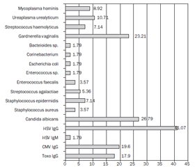
Актуальність. Проблема дефіциту вітаміну D у населення загалом і у вагітних зокрема та супутніх захворювань, у тому числі кістково-м’язової системи, залишається однією з найпоширеніших медико-соціальних проблем сучасності. Мета: визначити частоту захворювань кістково-м’язової системи у вагітних високого інфекційного ризику (ВІР) з порушеним статусом вітаміну D і однонуклеотидним Bsml поліморфізмом гена його рецепторів. Матеріали та методи. Обстежено 56 вагітних ВІР (основна група) та 40 практично здорових вагітних (контрольна група). Уміст 25-гідроксивітаміну D (25(ОН)D) у сироватці крові визначали методом імуноферментного аналізу; за допомогою методу полімеразної ланцюгової реакції у режимі реального часу виявляли мутантний варіант Bsml (rs1544410) поліморфізму гена, що кодує рецептори вітаміну D (VDR). Статистичну обробку результатів проводили за допомогою ресурсу www.socscistatistics.com. Результати. ВІР був зумовлений наявністю хронічних захворювань нирок, носійством збудників інфекцій групи TORCH та умовно-патогенної мікрофлори в урогенітальному тракті. Рівень 25(ОН)D був нижчим за загальноприйнятий оптимальний рівень у 76,8 і 15 % вагітних основної та контрольної груп відповідно (p = 0,0001). Носіями гетерозиготного генотипу A/G за геном VDR були 67,7 % вагітних ВІР проти 35 % у контрольній групі (відносний ризик (OR) = 3,95; 95% довірчий інтервал (ДІ): 2,19–7,1; χ2 = 20,88, p = 0,00001), а генотип G/G мали 19,6 і 47,5 % жінок відповідно (OR = 0,27; 95% ДІ 0,15–0,51; χ2 = 16,71, p = 0,00005). У третини вагітних з основної групи в анамнезі були захворювання кістково-м’язової системи (32,1 проти 12,5 %, OR = 3,15; 95% ДІ: 1,54–6,46), 71,4 % вагітних з цієї групи були носіями генотипу A/G (OR = 9,79; 95% ДI: 5,10–18,82). Висновки. Частка дефіциту й недостатності вітаміну D у вагітних ВІР становить майже 77 %. Загальносоматичний анамнез у цих жінок характеризується високою частотою захворювань нирок (37,5 %) і кістково-м’язової системи (32,1 %). Дві третини вагітних жінок ВІР, а також пацієнтки із захворюваннями кістково-м’язової системи є носіями гетерозиготного Bsml поліморфного генотипу A/G гена VDR, що, ймовірно, зумовлює більш високий ризик розвитку патології в умовах недостатності кальцитріолу. Вивчення статусу вітаміну D, генетична персоніфікація ризиків захворювань і своєчасна корекція модифікованих факторів, зокрема дефіциту вітаміну D, вважаються перспективними напрямками поліпшення перинатальних наслідків та якості життя вагітних.
Background. The problem of vitamin D (VD) deficiency in the population, in general, and in pregnant women, in particular, and related diseases, including the musculoskeletal system, remains one of the most widespread medical and social problems of our time. The purpose was to determine the frequency of musculoskeletal diseases in pregnant females at high infection risk (HIR) with impaired vitamin D status and single-nucleotide Bsml polymorphism of its receptor gene. Materials and methods. Fifty-six pregnant women (main group) with HIR and 40 healthy pregnant women (control group) had been examined. The level of 25-hydroxyvitamin D (25(OH)D) in blood was determined by the enzyme-linked immunosorbent assay, and real-time polymerase chain reaction was used to detect the mutant version of Bsml (rs1544410) polymorphism of the gene that is encoding vitamin D receptors (VDR). Statistical processing of the results was done using the resource www.socscistatistics.com. Results. HIR was due to the presence of chronic kidney diseases, carriers of pathogens of the TORCH group of infections and conditionally pathogenic microflora in the urogenital tract. The level of 25(OH)D was lower than the generally accepted optimal level in 76.8 and 15 % of pregnant women, in the main and control groups, respectively (F = 0.03; p = 0.0001). Carriers of the heterozygous genotype A/G were 67.7 % of pregnant women with HIR compared to 35 % of the control group (odd ratio (OR) = 3.95; 95% confidence interval (CI): 2.19–7.1; χ2 = 20.88, p = 0.00001), and the G/G genotype was inherent in 19.6 and 47.5 % of women, respectively (OR = 0.27; 95% CI 0.15–0.51; χ2 = 16.7, p = 0.00006). A third of pregnant women from the main group had a history of musculoskeletal diseases (32.14 %) versus 12.5 % in control group (OR = 3.15; 95% CI: 1.54–6.46); 71.4 % of pregnant women with HIR were carriers of A/G genotype (OR = 9.79; 95% CI: 5.10–18.82). Conclusions. The share of vitamin D deficiency/insufficiency in pregnant women with HIR is almost 77 %. The general somatic history of these women is characterized by a high frequency of the kidney diseases (37.5 %) and musculoskeletal diseases (32.1 %). Two-thirds of pregnant women with HIR, as well as with musculoskeletal diseases, are carriers of the heterozygous Bsml of polymorphic genotype A/G of the VDR gene, which probably causes a higher risk of the development of pathology in conditions of calcitriol deficiency. Studying VD status, the genetic personification of disease risks, and correction of modified factors in time, in particular, VD deficiency is seen as a promising direction for improving perinatal outcomes and the quality of life of pregnant women in general, but further research is required.
вагітність; Bsml поліморфізм гена рецепторів вітаміну D; інфікування; захворювання опорно-рухової системи
pregnancy; Bsml polymorphism of the vitamin D receptor gene; infection; musculoskeletal system diseases
Для ознакомления с полным содержанием статьи необходимо оформить подписку на журнал.
- Sebbag E., Felten R., Sagez F., Sibilia J., Devilliers H., Arnaud L. The world-wide burden of musculoskeletal diseases: a systematic analysis of the World Health Organization Burden of Diseases Database. Ann. Rheum. Dis. 2019 Jun. 78(6). 844-848. doi: 10.1136/annrheumdis-2019-215142.
- Clinton S., Newell A., Downey P., Ferreira K. Pelvic Girdle Pain in the Antepartum Population: Physical Therapy Clinical Practice Guidelines Linked to the International Classification of Functioning, Disability, and Health from the Section on Women’s Health and the Orthopaedic Section of the American Physical Therapy Association. Journal of Women’s Health Physical Therapy. 2017. 41(2). 102-125. doi: 10.1097/JWH.0000000000000081.
- Tankut V., Berenov K., Berenova O. Pelvic-spine pain at pregnancy: diagnostics and treatment. Orthopaedics, Traumatology and Prosthetics. 2020. 3. 61-66. doi: 10.15674/0030-59872020361-66.
- Bortkevich O.P., Bilyavska Yu.V., Korendovych V.V. Updated approaches to the treatment of rheumatic disea–ses in pregnant and lactating women. Ukrainian Journal of Rheumatology. 2016. 64(2). 25-33. Available from: https://www.rheumatology.kiev.ua/article/8958/onovleni-pidxodi-do-likuvannya-revmatichnix-xvorob-u-zhinok-v-period-vagitnosti-ta-laktacii.
- Zhu X., Chan Y.T., Yung P.S.H., Tuan R.S., Jiang Y. Subchondral Bone Remodeling: A Therapeutic Target for Osteoarthritis. Front. Cell Dev. Biol. 2021 Jan 21. 8. 607764. doi: 10.3389/fcell.2020.607764.
- Schmitt S. Chronic Infectious Arthritis. 2022. Avai–lable from: https://www.msdmanuals.com/professional/musculoskeletal-and-connective-tissue-disorders/infections-of-joints-and-bones/acute-infectious-arthritis.
- Vitamin D: screening and supplementation during pregnancy. Committee Opinion No. 495. American College of Obstetricians and Gynecologists. Obstet. Gynecol. 2011. 118. 197-8. Available from: https://www.acog.org/clinical/clinical-guidance/committee-opinion/articles/2011/07/vitamin-d-screening-and-supplementation-during-pregnancy.
- Danese E., Pucci M., Montagnana M., Lippi G. Vitamin D deficiency and pregnancy disorders. Journal of Laboratory and Precision Medicine. 2019. 5. doi: 10.21037/jlpm.2019.11.03.
- Khattab Y., Reda R., El-Gaafary M. et al. BsmI gene polymorphism of vitamin D receptor in obese Egyptian male medical students and its relationship with vitamin D deficiency. Egypt. J. Med. Hum. Genet. 2022. 23. doi: 10.1186/s43042-022-00275-z.
- Palacios C., Kostiuk L.K., Peña-Rosas J.P. Vitamin D supplementation for women during pregnancy. Cochrane Database of Systematic Reviews. 2019. 7. CD008873. doi: 10.1002/14651858.CD008873.pub4.
- Bharti B., Lee S.J., Lindsay S.P., Wingard D.L., Jones K.L., Lemus H., Chambers C.D. Disease Seve–rity and Pregnancy Outcomes in Women with Rheumatoid Arthritis: Results from the Organization of Teratology Information Specialists Autoimmune Diseases in Pregnancy Project. J. Rheumatol. 2015 Aug. 42(8). 1376-82. doi: 10.3899/jrheum.140583.
- Qureshi S., Kanzali M., Rizvi S.F., Joolukuntla N., Fomberstein B. New diagnosis of rheumatoid arthritis during the third trimester of pregnancy. Women’s Health (Lond.). 2016 Jul. 12(4). 407-11. doi: 10.1177/1745505716661724.
- Yucesoy B., Charles L.E., Baker B., Burchfiel C.M. Occupational and genetic risk factors for osteoarthritis: a review. Work. 2015 Jan 1. 50(2). 261-73. doi: 10.3233/WOR-131739.
- Li Y., Zhu J., Gao C., Peng B. Vitamin D receptor (VDR) genetic polymorphisms associated with intervertebral disc degeneration. J. Genet. Genomics. 2015 Apr 20. 42(4). 135-40. doi: 10.1016/j.jgg.2015.03.006.
- Colombini A., Brayda-Bruno M., Lombardi G., Croiset S.J., Ceriani C. et al. BsmI, ApaI and TaqI Polymorphisms in the Vitamin D Receptor Gene (VDR) and Association with Lumbar Spine Pathologies: An Italian Case-Control Study. PLoS One. 2016 May 5. 11(5). e0155004. doi: 10.1371/journal.pone.0155004.
- Luo L., Li X., Yan R., Zhang H., Li C. Risk factors for adverse pregnancy outcomes in women with rheumatoid arthritis and follow-up of their offspring. Clinical Rheumatology. 2022 Oct. 41(10). 3135-3141. doi: 10.1007/s10067-022-06233-9.
- Kesikburun S., Güzelküçük Ü., Fidan U., Demir Y., Ergün A., Tan A.K. Musculoskeletal pain and symptoms in pregnancy: a descriptive study. Ther. Adv. Musculoskelet. Dis. 2018 Nov 19. 10(12). 229-234. doi: 10.1177/1759720X18812449.
- Mathew A.J., Ravindran V. Infections and arthritis. Best Pract. Res. Clin. Rheumatol. 2014 Dec. 28(6). 935-59. doi: 10.1016/j.berh.2015.04.009.
- Sinyachenko O.V., Yermolaeva M.V., Liventsova K.V., Verzilov S.M., Alieva T.Yu., Haviley D.O. Rheumatoid arthritis and comorbid periodontitis: clinicopathogenetic features of the relationship. Ukrainian Journal of Rheumatology. 2020. 82(4). 4-11. doi: 10.32471/rheumatology.2707-6970.82.15656 (in Ukrainian).
- Fabbri A., Infante M., Ricordi C. Editorial — Vitamin D status: a key modulator of innate immunity and natural defense from acute viral respiratory infections. Eur. Rev. Med. Pharmacol. Sci. 2020 Apr. 24(7). 4048-4052. doi: 10.26355/eurrev_202004_20876.
- Saraf R., Morton S.M., Camargo C.A. Jr, Grant C.C. Global summary of maternal and newborn vitamin D status — a systematic review. Matern. Child Nutr. 2016 Oct. 12(4). 647-68. doi: 10.1111/mcn.12210.
- Ince-Askan H., Hazes J.M.W., Dolhain R.J.E.M. Identifying clinical factors associated with low disease acti–vity and remission of rheumatoid arthritis during pregnancy. Arthritis Care Res. (Hoboken). 2017 Sep. 69(9). 1297-1303. doi: 10.1002/acr.23143.

