Международный эндокринологический журнал Том 20, №4, 2024
Вернуться к номеру
Рання діагностика мінеральних і кісткових розладів у пацієнтів iз діабетичною хворобою нирок на тлі цукрового діабету 2-го типу
Авторы: V.M. Yerokhovych (1), O.V. Karpenko (1), I.A. Paliienko (1), N.M. Kobyliak (1), M.I. Bobryk (1), L.V. Shuliarenko (1), O.A. Rudenko (1), D.V. Kyriienko (2), M. Bolanowski (3), Y.I. Komisarenko (1)
(1) - Bogomolets National Medical University, Kyiv, Ukraine
(2) - Kyiv City Clinical Endocrinology Center, Kyiv, Ukraine
(3) - Wrocław Medical University, Wrocław, Poland
Рубрики: Эндокринология
Разделы: Клинические исследования
Версия для печати
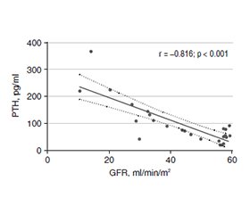
Актуальність. Цукровий діабет є актуальною проблемою сьогодення, яка характеризується прогресуючим зростанням кількості пацієнтів із високою частотою ускладнень, що потребують ранньої діагностики та своєчасних лікувальних заходів. Одним із найпоширеніших мікросудинних уражень є діабетична нефропатія. Пацієнти можуть мати клінічні прояви діабетичної хвороби нирок, які виходять за межі класичних симптомів і мають екстраренальні наслідки у вигляді мінерало-кісткових розладів. Мета роботи: провести комплексну оцінку ранніх маркерів ураження нирок і змін показників мінеральної щільності кісткової тканини в пацієнтів із цукровим діабетом 2-го типу, а також виявити взаємозв’язки досліджуваних параметрів. Матеріали та методи. У дослідженні взяли участь 80 пацієнтів із цукровим діабетом 2-го типу, які були розподілені за рівнем швидкості клубочкової фільтрації: рШКФ < 60 мл/хв/м2 (перша група, n = 26), рШКФ ≥ 60 мл/хв/м2 (друга група, n = 54). Результати. Аналіз ранніх маркерів ураження нирок в обстежених групах виявив деякі суттєві відмінності. Показники співвідношення альбуміну та креатиніну в добовій сечі, сироваткового цистатину С, паратгормону, сечової кислоти, вітамін-D-зв’язуючого білка були вірогідно вищими в пацієнтів із рШКФ < 60 мл/хв/м2. Середній уміст вітаміну D в обох групах відповідав дефіцитному стану, причому перша група відзначалася статистично вірогідно нижчим рівнем порівняно з другою — 12,32 ± 4,84 та 16,72 ± 5,82 нг/мл відповідно (р = 0,001). У першій групі дефіцит вітаміну D спостерігався в 92,3 % випадків, у другій — у 74,1 % (р = 0,56). При кореляційному аналізі знайдені деякі вірогідні зв’язки: у першій групі — негативна кореляція між рШКФ та ПТГ (r = –0,816, р < 0,001). Спостерігався обернений зв’язок між рШКФ та цистатином С у 1-й (r = –0,862, p < 0,001) та 2-й групах (r = –0,322, p = 0,18). Серед усіх обстежених учасників виявлено лінійну негативну кореляцію між рШКФ та рівнем сечової кислоти (r = –0,452; p < 0,001). Вітамін D не мав вірогідного зв’язку з рШКФ, проте ми знайшли негативну кореляцію зі співвідношенням альбуміну й креатиніну в добовій сечі (r = –0,253, р = 0,024) та цистатином С (r = –0,303, p = 0,006), що підтверджує роль холекальциферолу в порушенні мінеральної щільності кісткової тканини в пацієнтів із діабетичною хворобою нирок. У нашому дослідженні виявлено зворотну кореляцію між рШКФ та вітамін-D-зв’язуючим білком у першій (r = –0,436, р = 0,26) та другій групі (r = –0,283, р = 0,038), що, ймовірно, вказує на можливу компенсаторну реакцію транспортного білка на початкові мінерало-кісткові порушення в пацієнтів із діабетичним ураженням нирок. Висновки. Раннє виявлення мінеральних і кісткових розладів при діабетичній хворобі нирок є важливим щодо підвищення ефективності ведення пацієнтів із цукровим діабетом 2-го типу та своєчасного лікування, профілактики ускладнень з боку серцево-судинної системи й порушень кісткового метаболізму.
Background. Today, diabetes mellitus is an actual problem, characterized by a progressive increase in the number of patients with a high frequency of complications that require early diagnosis and timely treatment. Diabetic nephropathy is among the most common microvascular lesions. Patients may have clinical manifestations of diabetic kidney disease that go beyond the classic symptoms and have extrarenal consequences in the form of bone mineral disorders. The purpose of the work is to carry out a comprehensive assessment of early markers of kidney damage and changes in bone disorder indicators in patients with type 2 diabetes and to identify correlations between the studied parameters. Materials and methods. Eighty patients with type 2 diabetes participated in the study. They were divided according to the glomerular filtration rate: GFR < 60 ml/min/m2 (1st group, n = 26), GFR ≥ 60 ml/min/m2 (2nd group, n = 54). Results. Analysis of early markers of kidney damage revealed some significant differences between the groups. Indicators of daily urine albumin-creatinine ratio, serum cystatin C, parathyroid hormone, uric acid, and vitamin D-binding protein were significantly higher in patients with GFR < 60 ml/min/m2. The average level of vitamin D (25OH) in both groups corresponded to a deficient state, and the 1st group was marked by a statistically significantly lower level compared to the 2nd group: 12.32 ± 4.84 and 16.72 ± 5.82 ng/ml, respectively (p = 0.001). In the 1st group, vitamin D deficiency was observed in 92.3 % of cases, and in the 2nd group, in 74.1 % (p = 0.56). According to the correlation analysis, some reliable relationships were found: in the 1st group, there was a negative correlation between GFR and parathyroid hormone (r = –0.816, p < 0.001). An inverse correlation was revealed between GFR and cystatin C in the 1st (r = –0.862, p < 0.001) and 2nd groups (r = –0.322, p = 0.18). Among all examined participants, there was a linear negative correlation between GFR and uric acid (r = –0.452, p < 0.001). Vitamin D (25OH) didn’t have a significant relationship with GFR, however, we found a negative correlation with the daily urine albumin-creatinine ratio (r = –0.253, р = 0.024) and cystatin C (r = –0.303, p = 0.006), which confirms the role of cholecalciferol in mineral bone disorders in patients with chronic kidney disease. In our study, an inverse correlation was found between GFR and vitamin D-binding protein in the 1st (r = –0.436, p = 0.26) and 2nd group (r = –0.283, p = 0.038), which probably indicates a possible compensatory response of transport protein to initial mineral bone disorders in patients with diabetic kidney disease. Conclusions. Early detection of bone mineral disorders in diabetic kidney disease is important to increase the efficiency of managing patients with type 2 diabetes and timely treatment, prevention of cardiovascular complications and bone metabolism disorders.
цукровий діабет; діабетична хвороба нирок; хронічна хвороба нирок; дефіцит вітаміну D; мінеральні та кісткові розлади; гіперпаратиреоз; альбумінурія; вітамін-D-зв’язуючий білок
diabetes mellitus; diabetic kidney disease; chronic kidney disease; vitamin D deficiency; mineral bone disorders; hyperparathyroidism; albuminuria; vitamin D-binding protein
Для ознакомления с полным содержанием статьи необходимо оформить подписку на журнал.
- Sun H, Saeedi P, Karuranga S, Pinkepank M, Ogurtsova K, et al. IDF Diabetes Atlas: Global, regional and country-level diabetes prevalence estimates for 2021 and projections for 2045. Diabetes Res Clin Pract. 2022 Jan;183:109119. doi: 10.1016/j.diabres.2021.109119.
- Thipsawat S. Early detection of diabetic nephropathy in patient with type 2 diabetes mellitus: A review of the literature. Diab Vasc Dis Res. 2021 Nov-Dec;18(6):14791641211058856. doi: 10.1177/14791641211058856.
- Yu SM, Bonventre JV. Acute Kidney Injury and Progression of Diabetic Kidney Disease. Adv Chronic Kidney Dis. 2018 Mar;25(2):166-180. doi: 10.1053/j.ackd.2017.12.005.
- Ke Y, Jian-Yuan H, Ping Z, Yue W, Na X, et al. The progressive application of single-cell RNA sequencing technology in cardiovascular diseases. Biomed Pharmacother. 2022 Oct;154:113604. doi: 10.1016/j.biopha.2022.113604.
- Pankiv I. The prevalence and structure of major risk factors in women with postmenopausal osteoporosis. International Journal of Endocrinology (Ukraine). 2018;14(8):744-748. doi: 10.22141/2224-0721.14.8.2018.154854.
- Pereira PR, Carrageta DF, Oliveira PF, Rodrigues A, Alves MG, Monteiro MP. Metabolomics as a tool for the early diagnosis and prognosis of diabetic kidney disease. Med Res Rev. 2022 Jul;42(4):1518-1544. doi: 10.1002/med.21883.
- Hu L, Napoletano A, Provenzano M, Garofalo C, Bini C, et al. Mineral Bone Disorders in Kidney Disease Patients: The Ever-Current Topic. Int J Mol Sci. 2022 Oct 13;23(20):12223. doi: 10.3390/ijms232012223.
- Chapter 4: Other complications of CKD: CVD, medication dosage, patient safety, infections, hospitalizations, and caveats for investigating complications of CKD. Kidney Int Suppl (2011). 2013 Jan;3(1):91-111. doi: 10.1038/kisup.2012.67.
- Pazianas M, Miller PD. Osteoporosis and Chronic Kidney Disease-Mineral and Bone Disorder (CKD-MBD): Back to Basics. Am J Kidney Dis. 2021 Oct;78(4):582-589. doi: 10.1053/j.ajkd.2020.12.024.
- Yamada S, Nakano T. Role of Chronic Kidney Disease (CKD)-Mineral and Bone Disorder (MBD) in the Pathogenesis of Cardiovascular Disease in CKD. J Atheroscler Thromb. 2023 Aug 1;30(8):835-850. doi: 10.5551/jat.RV22006.
- Grygorieva N, Tronko M, Kovalenko V, Komisarenko S, Tatarchuk T, et al. Ukrainian Consensus on Diagnosis and Mana–gement of Vitamin D Deficiency in Adults. Nutrients. 2024 Jan 16;16(2):270. doi: 10.3390/nu16020270.
- Feng J, Wang H, Jing Z, Wang Y, Cheng Y, et al. Role of Magnesium in Type 2 Diabetes Mellitus. Biol Trace Elem Res. 2020 Jul;196(1):74-85. doi: 10.1007/s12011-019-01922-0.
- Hughson MD, McCarty GA, Sholer CM, Brumback RA. Thrombotic cerebral arteriopathy in patients with the antiphospholipid syndrome. Mod Pathol. 1993 Nov;6(6):644-53.
- Bai X, Luo Q, Tan K, Guo L. Diagnostic value of VDBP and miR-155-5p in diabetic nephropathy and the correlation with urinary microalbumin. Exp Ther Med. 2020 Nov;20(5):86. doi: 10.3892/etm.2020.9214.
- Kidney Disease: Improving Global Outcomes (KDIGO) Diabetes Work Group. KDIGO 2022 Clinical Practice Guideline for Diabetes Management in Chronic Kidney Disease. Kidney Int. 2022 Nov;102(5S):S1-S127. doi: 10.1016/j.kint.2022.06.008.
- Haines RW, Fowler AJ, Liang K, Pearse RM, Larsson AO, et al. Comparison of Cystatin C and Creatinine in the Assessment of Measured Kidney Function during Critical Illness. Clin J Am Soc Nephrol. 2023 Aug 1;18(8):997-1005. doi: 10.2215/CJN.0000000000000203.
- Bobryk M, Tutchenko T, Sidorova I, Burka O, Krotyk O, Serbeniuk A. Insulin resistance in the ХХІ century: multimodal approach to assessing causes and effective correction. Reproductive Endocrinology. 2021;62:97-103. doi: 10.18370/2309-4117.2021.62.97-103.
- Rodríguez-Ortiz ME, Rodríguez M. Recent advances in understanding and managing secondary hyperparathyroidism in chronic kidney disease. F1000Res. 2020 Sep 1;9:F1000 Faculty Rev-1077. doi: 10.12688/f1000research.22636.1.
- Yerokhovych V, Komisarenko Y, Karpenko O, Pankiv V, Kobyliak N, et al. Assessment of renal and cardiovascular risks in patients with type 2 diabetes when using non-steroidal mineralocorticoid receptor antagonists. International Journal of Endocrinology (Ukraine). 2024;19(8):579-585. doi: 10.22141/2224-0721.19.8.2023.1341.

